Diagnostic methods and techniques for Diabetic Retinopathy

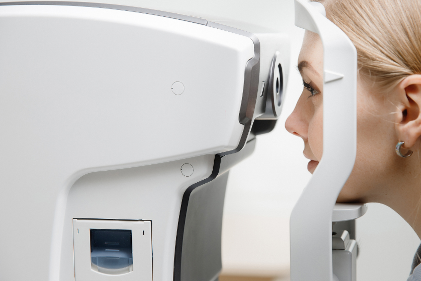
Die Optische Kohärenztomographie (OCT) ist eine nicht-invasive Methode zur Diabetischen Retinopathie Diagnose
In our day-to-day practice, it is unfortunately confirmed almost daily: Diabetic Retinopathy (DR) is a serious eye disease that requires early diagnosis and treatment in order to protect vision.
Doctors use various diagnostic methods and techniques to assess the condition of the retina and identify changes:
Eye examination: a direct view of the retina
The eye examination is the essential first step in the diagnosis of Diabetic Retinopathy. This procedure allows doctors to delve directly into the fine structure of the retina to identify possible changes.
This makes it possible to better assess the severity of the disease and make an informed treatment decision to best protect the patient’s vision.
Regular performance of these examinations is crucial to ensure timely intervention.
Two key methods are used in the diagnosis of Diabetic Retinopathy:
Ophthalmoscopy as a method of eye examination
This is a technique in which the ophthalmologist uses an ophthalmoscope, a special eye examination device, to check the retina for abnormalities and deviations.
The patient’s pupils are dilated to enable the doctor to view the inside of the eye. This method makes it possible to detect signs of haemorrhages, exudates (secretions of fluids) and neovascularisation (abnormal vascular formations).
In addition, the condition of the blood vessels can be analysed to obtain indications of circulatory disorders.
Slit lamp examination as a method of eye examination
This procedure expands the doctor’s field of vision and allows him to take a closer look at the front and back of the eye. A narrow band of light is directed at the patient’s eye while the doctor looks at the structures with a magnifying glass.
This allows the condition of the lens, the cornea, the anterior segment of the eye and the retina to be assessed more accurately. This method is particularly helpful in recognising possible damage to the optic nerve, which plays a key role in the visual process.
The eye examination serves as an important basis for the early detection of Diabetic Retinopathy, as it enables doctors to recognise not only changes in the retina, but also possible abnormalities in the eye as a whole.
Fundus photography: detailed insights into retinal changes
Fundus photography, a crucial method in the diagnosis of Diabetic Retinopathy, allows doctors to take high-resolution images of the retina, providing a precise image of changes.
This procedure allows doctors to analyse microscopic abnormalities such as microaneurysms, haemorrhages and swellings in detail.
By visually documenting the progression of the disease over time, doctors can track changes at different stages of Diabetic Retinopathy and adjust treatment plans accordingly.
Fundus photography thus plays an essential role in assessing disease progression and accurately monitoring eye health in patients with Diabetic Retinopathy.
Optical coherence tomography (OCT): Precise insights into the retinal layers
Optical Coherence Tomography (OCT) is a sophisticated imaging technique that produces an impressive layer-by-layer visualisation of the retina.
This non-invasive method allows doctors to analyse the structure of the retina in detail by measuring the thickness of the individual layers. This precise measurement allows medical professionals to identify changes such as macular oedema, i.e. swelling in the area of sharpest vision.
This procedure not only enables the early detection of potential complications, but also allows an accurate assessment of the severity of Diabetic Retinopathy.
OCT therefore provides valuable insights into the microscopic changes in the retina and supports the medical assessment and targeted management of this eye disease.
Fluorescein angiography: detailed insights into the blood flow and vascular structure of the retina
Fluorescein angiography is a diagnostic procedure in which a contrast agent is injected into the patient’s bloodstream. This contrast agent flows through the blood vessels of the retina and enables doctors to visualise the blood flow and vascular structure in high resolution.
This technique allows the detection of leaks, abnormal vessels and circulatory disorders often associated with Diabetic Retinopathy. In particular, doctors can identify neovascularisation, i.e. the formation of new, unwanted blood vessels.
The information obtained from fluorescein angiography helps to accurately assess the progression of the disease and plan suitable treatment approaches.
Visual field test: recognising damage in the peripheral field of vision
The visual field test is an important method for assessing visual function and the peripheral field of vision in patients with Diabetic Retinopathy. This test allows doctors to recognise changes in the visual field that indicate damage to the retina.
During the test, the patient looks at a fixed point in the centre of the screen and presses a button as soon as they notice a light signal in the peripheral field of vision.
The results of the visual field test allow doctors to identify narrowing or loss in the field of vision, often caused by damage to the retina.
This test is crucial to understand the potential impact of Diabetic Retinopathy on vision and to adjust the treatment strategy accordingly.
A regular visual field test helps to monitor changes in vision over time and to react early to potential complications.
Electroretinogram (ERG): A functional analysis of the retinal cells
The electroretinogram (ERG) is an advanced technique for Diabetic Retinopathy diagnosis that provides insight into the electrical activity of retinal cells in Diabetic Retinopathy.
Using electrodes placed on the surface of the eye, doctors can measure the retina’s response to light stimuli.
This method makes it possible to recognise functional disorders of the retina at an early stage and determine the degree of damage. The ERG provides valuable information about visual function and helps to assess the condition of the retinal cells in Diabetic Retinopathy.
During the ERG test, the patient is presented with a series of light stimuli while the electrodes record the electrical signals from the retina.
The data obtained is analysed to identify deviations from normal retinal function in Diabetic Retinopathy.
The ERG is particularly useful in the assessment of this eye disease, where retinal cell function may be impaired. The results of the ERG provide doctors with important information about the condition of the retina.
The ERG is therefore an important pillar of Diabetic Retinopathy diagnosis.
Diabetic Retinopathy Diagnosis: Total body examination for comprehensive control
Diabetic Retinopathy is closely linked to the cause of diabetes as a systemic disease. It is therefore crucial to keep an eye not only on the eyes, but also on the patient’s entire body.
A comprehensive total body examination can help to slow down the progression of Diabetic Retinopathy and minimise the risk of complications.
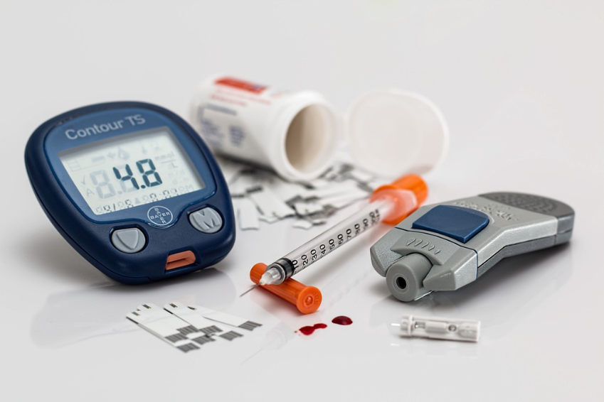
Controlling blood glucose levels and blood pressure plays a central role in the management of Diabetic Retinopathy and Diabetic Retinopathy diagnosis.
Optimal blood glucose control can help reduce the risk of damage to the blood vessels of the retina and thus influence the development of Diabetic Retinopathy.
At the same time, good blood pressure control can help to protect the vessels in the retina and minimise the risk of circulatory disorders.
Close co-operation with the family doctor, ophthalmologist and other specialists is of great importance.
Coordinating the treatment of diabetes and its associated conditions can help to improve overall health and reduce the risk of complications, including Diabetic Retinopathy.
Regular monitoring of other risk factors such as elevated blood lipid levels and kidney function is also important to develop a holistic treatment strategy.

