Glaucoma treatment – a complete overview of all treatment methods

Glaucoma treatment - we give a complete overview of all Glaucoma treatment methods.

Glaucoma treatment – we give a complete overview of all Glaucoma treatment methods.

Effective treatment of glaucoma is essential to slow or stop the progressive loss of vision. It is important to understand that glaucoma cannot be cured as damage already done to the optic nerve is irreversible.

These methods of glaucoma treatment are available and are described in detail:

The main method of lowering intraocular pressure in glaucoma is the use of special eye drops. These preparations are applied once a day or several times a day, depending on the prescription. Regular check-ups at the ophthalmologist’s office are also of great importance and are usually performed every 1 to 3 months, depending on the progress of the disease.

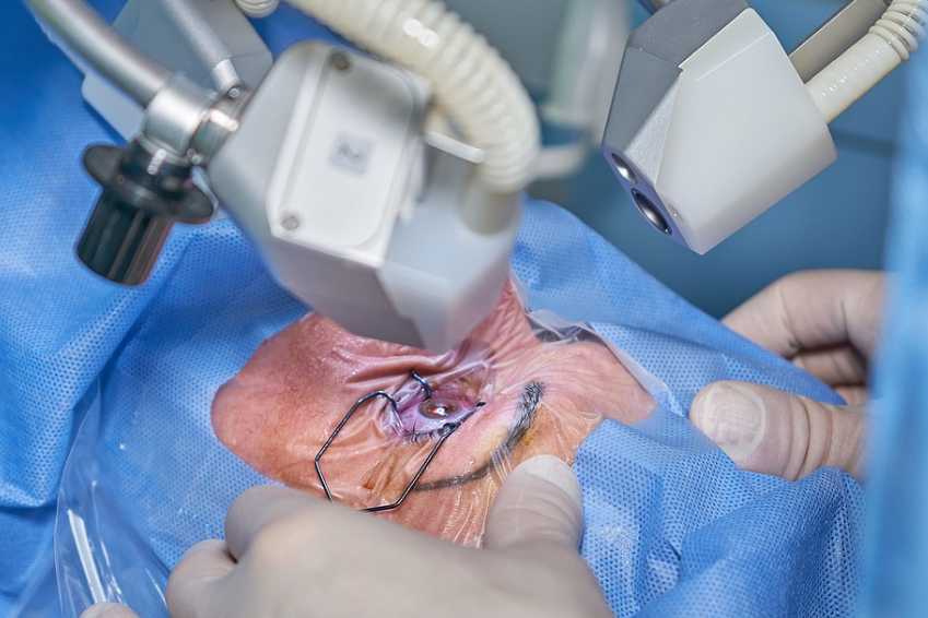
In cases where drug treatment is not sufficiently effective or is not well tolerated, surgical procedures such as trabeculectomy or minimally invasive procedures may be considered.

In the event of an acute glaucoma attack, it is imperative to seek medical attention immediately to prevent damage to the optic nerve. In such emergencies, laser treatment can help to quickly reduce the congested intraocular pressure.

Therapy for glaucoma includes:

  • Application of medication in the form of eye drops
  • Laser interventions
  • Surgical procedures

The main aim of these treatments is to lower the intraocular pressure to minimise the risk of vision damage and blindness.

The term glaucoma, refers to several eye diseases that all result in damage to the optic nerve. The most common glaucoma symptoms include:

The reduction of intraocular pressure is crucial to slow down or stop the gradual loss of vision in glaucoma. Nevertheless, it is important to emphasise that a cure for glaucoma is not possible, as existing damage to the optic nerve cannot be reversed.

In the following, we describe in full breadth and depth:

  • The basic treatment options for each type of glaucoma
  • The options for treating glaucoma in children
  • The medicines used for glaucoma treatment
  • The alternative treatment options
  • New therapies for glaucoma treatment
  • The different procedures for glaucoma surgery
  • As a subgroup of surgeries, the different laser surgery procedures
  • The glaucoma cure options

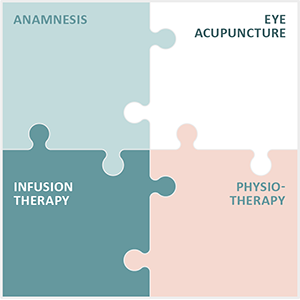
Integrated Eye Therapy according to Noll

Augenakupunktur Noll specialises in the treatment of degenerative eye diseases.

Read more

The Pseudoexfoliation Syndrome treatment

Pseudoexfoliative glaucoma syndrome

Pseudoexfoliative glaucoma, also known as pseudoexfoliative glaucoma, is a common complication of pseudoexfoliation syndrome. PEX glaucoma is a type of secondary open-angle glaucoma with increased intraocular pressure and has a higher rate of progression compared to other forms of glaucoma, meaning that it carries a greater chance of optic nerve damage.

There are some differences between PEX glaucoma and primary open-angle glaucoma. PEX glaucoma is characterised by more pigmentation in the chamber angle and higher intraocular pressures. It tends to cause pressure spikes and fluctuations throughout the day, as well as acute pressure increases associated with pupil dilation.

In addition, optic nerve damage and visual field loss occur more quickly and frequently in PEX glaucoma. These differences in symptoms are also associated with greater resistance to drug therapy.

About half of people with pseudoexfoliation syndrome also develop pseudoexfoliation glaucoma over time, making PEX syndrome one of the most common causes of glaucoma overall. About one-third of patients diagnosed with this form of glaucoma have the condition in both eyes.

The deposits that occur with pseudoexfoliation syndrome in the lens of the eye and in the chamber angle cause the chamber angle to close. This in turn prevents the normal outflow of aqueous humour and leads to an increase in intraocular pressure. With this increased pressure on the optic nerve, the risk of damage increases, eventually leading to the development of glaucoma.

In advanced stages, pseudoexfoliative glaucoma, like all glaucomas, causes visual field loss. Compared to primary open-angle glaucoma, PEX glaucoma progresses more rapidly and responds less well to drug treatments. Therefore, surgical intervention is necessary in many cases.

Causes for Pseudoexfoliation Syndrome

The exact causes for the occurrence of pseudoexfoliation syndrome still remain unclear in research. However, there are scientific studies that suggest several possibilities:

One possible cause could be certain inflammatory reactions and processes that could favour the development of pseudoexfoliation syndrome. On the other hand, studies suggest that genetic factors as well as oxidative stress could play a significant role.

There is also evidence that elevated homocysteine levels could favour the occurrence of pseudoexfoliation syndrome. Homocysteine is an amino acid of interest in the context of cardiovascular disease, particularly cardiovascular disease.

Furthermore, studies show that pseudoexfoliation syndrome is more common in patients with mutations in the LOXL1 gene. This gene plays a crucial role in elastin metabolism, and mutations can apparently lead to the production of the pseudoexfoliative material.

However, it is important to emphasise that the presence of this gene defect is not the only factor contributing to the development of pseudoexfoliation syndrome, and it does not necessarily lead to the development of glaucoma.

Another significant factor in the development of pseudoexfoliation syndrome is age. The risk of developing this condition increases with age, especially in people over 70. In Switzerland, about one in 20 people is affected, with women having a higher risk.

In view of the ageing population, the syndrome is becoming increasingly clinically important in both women and men.

Diagnosis of Pseudoexfoliation Syndrome (PEX): recognition and criteria

In advanced pseudoexfoliation syndrome, the ophthalmologist can identify specific abnormal findings. For example, the slit lamp can be used to identify proteinaceous accumulations at the edge of the pupil and deposits on the lens of the eye.

In addition, an examination of the chamber angle, known as gonioscopy, can detect deposits of PEX material in the dense tissue of the chamber angle. These deposits can obstruct the natural outflow of aqueous humour.

The PEX material, which is formed in various parts of the eye both inside and outside, is made up of fine fibrous elastic components derived from various cells and tissues. In the eye, whitish, flaky particles are visible on the lens and its inner edge, as well as on the iris.

Diagnosis of pseudoexfoliation syndrome can be challenging in many cases due to the absence of central deposits. A reliable diagnosis is often only possible after pupil dilation.

In the early stages, the presence of the disease can be detected by factors such as subtle changes in the lens combined with pigment dispersion (detachment of pigment cells from the back of the iris) or mydriasis weakness (malfunction in pupil dilation).

Because of the high risk of developing glaucoma, early diagnosis of PEX syndrome is highly desirable. Accurate and early diagnosis is particularly clinically relevant, especially when considering the potential need for cataract surgery (lens replacement surgery) as part of cataract treatment and the associated risks.

Clinical criteria for the diagnosis of Pseudoexfoliation Syndrome

The clinical criteria for the diagnosis of pseudoexfoliation syndrome include:

  • Disc-shaped deposits: of grey, scaly material on the front of the lens and in the chamber angle.
  • Iris flutter: which is manifested by the trembling of the iris due to poor adherence to the environment, for example during head movements.
  • Lens luxation: where the lens is displaced into the anterior chamber of the eye or into the vitreous cavity.
  • Rubeosis Iridis: a pinkish-red colouration of the iris or increased vascularisation in the iris and the chamber angle.
  • Secondary optic atrophy: which refers to the loss of nerve cells of the optic nerve.

Symptoms of Pseudoexfoliation Syndrome

Often, pseudoexfoliation syndrome has no obvious symptoms and does not cause any pain. However, if complications such as glaucoma develop, symptoms may develop that can lead to damage to the optic nerve head and progressive vision loss.

But what risk factors increase the likelihood of developing pseudoexfoliation syndrome?

The risk for this condition increases significantly with age, especially in people over 70 years of age. In the general population, on average, about one in 20 people is affected, with women having a higher risk of developing the disease. With the steady increase in the average age of the population, the prevalence of pseudoexfoliation syndrome is also increasing.

In addition, genetic predispositions play an important role. Individuals who have a family history of the condition are at increased risk. Similarly, residence in northern countries may increase the risk for pseudoexfoliation syndrome.

Overall, it is crucial to keep these risk factors in mind and have regular ophthalmological examinations to detect possible symptoms and signs early and take appropriate measures for prevention and treatment.

Treatment options for Pseudoexfoliation Syndrome

Since the exact causes of pseudoexfoliation syndrome cannot be treated or are not yet fully known, therapy focuses on preventing further damage to the eye.

For example, if the intraocular pressure is elevated, attempts can be made to lower it with eye drops to reduce the risk of developing glaucoma. There are different types of eye drops available that are suitable for reducing intraocular pressure.

Usually, treatment starts with an eye drop preparation that contains only one active ingredient. If the reduction of the intraocular pressure is not sufficient, the combination of several active substances can be considered. However, usually no more than two different eye drop preparations with a total of three different active ingredients are used.

If there is a blocked chamber angle, an attempt can be made to open it up using a laser to improve the drainage of the aqueous humour in the trabecular meshwork. This procedure, known as Selective Laser Trabeculoplasty, treats the trabecular meshwork with a laser beam, which causes the pores in the tissue to enlarge and improves outflow.

Selective laser trabeculoplasty is a gentle procedure and can be used at the beginning of glaucoma therapy or in addition to the use of eye drops. If the effect of the laser procedure wears off, the treatment can be repeated without any problems.

Normally, laser treatment can reduce intraocular pressure by about 30 percent. However, this effect often does not last very long and the need for surgical intervention occurs more quickly than with other forms of glaucoma.

Finally, if the intraocular pressure cannot be lowered sufficiently, the only option is to surgically remove the lens, which is known as cataract surgery. Removing the lens offers the possibility of eliminating the cause of the pseudoexfoliation syndrome, which can lead to a normalisation of the intraocular pressure after the operation. Especially in cases of severe pressure fluctuations and simultaneous presence of a cataract, a surgical approach should be considered.

Standard procedures for surgical therapy

These are the standard procedures for the surgical treatment of pseudoexfoliation syndrome at a glance:

  • Trabeculectomy (filter pad surgery): This involves preparing a small hole in the wall of the eyeball, which is covered with part of the sclera. This allows the aqueous humour to drain under the conjunctiva when the intraocular pressure is high, where a kind of seepage cushion is formed.
  • Canaloplasty (thread surgery): This technique widens Schlemm’s canal with the help of a microcatheter and a special gel and stretches a fine thread into the canal. This improves eye drainage without opening the eyeball itself. However, canaloplasty does not usually achieve the same pressure reduction as trabeculectomy, which is why additional eye drops are often required.
  • iStent implant: In this minimally invasive procedure, two titanium implants are inserted into the chamber angle in the trabecular meshwork. This allows the aqueous humour to flow directly into Schlemm’s canal. The implantation can be done alone or at the same time as cataract surgery to further reduce intraocular pressure.
  • XEN gel implant: This is a minimally invasive procedure in which a gelatin tube is inserted into the anterior chamber to create a seepage cushion, similar to trabeculectomy.
  • Drainage implants: If medication and trabeculectomy do not adequately lower intraocular pressure, a silicone tube may be inserted into the anterior chamber to allow fluid drainage.
  • Cyclophotocoagulation: This procedure uses a laser to sclerotise the ciliary body, which is responsible for producing the eye’s fluid. This reduces the amount of fluid produced and lowers the intraocular pressure.

It should be noted that there are risks after both laser treatments and surgical procedures in the early post-operative phase. This is because inflammatory reactions and pressure spikes can occur when the blood-chamber fluid barrier breaks down.

These reactions occur frequently, so intensive monitoring and treatment during and after the procedures is of great importance.

It is also important to note that the therapy for pseudoexfoliation syndrome is essentially the same as that for primary open-angle glaucoma. However, it is crucial to change the therapeutic approach more quickly and consistently from conservative to surgical measures.

Prognosis of Pseudoexfoliation Syndrome

The prognosis for pseudoexfoliation syndrome and the glaucoma that is often associated with it depends crucially on timely detection. Thanks to a wide range of customisable treatment options, there is now a chance of a favourable prognosis. However, if left untreated, the syndrome can lead to premature blindness.

Comparing the symptoms of patients diagnosed with pseudoexfoliative glaucoma with those of patients with primary chronic open-angle glaucoma at the time of detection shows that the average intraocular pressure is significantly higher in pseudoexfoliative glaucoma patients.

In addition, about a quarter of pseudoexfoliative glaucoma patients are virtually blind in the affected eyes, while only about 15 per cent of primary open-angle glaucoma sufferers show a slight deterioration in vision.

Furthermore, those affected by the pseudoexfoliation syndrome show deposits in the chamber angle significantly more often. Therefore, regular ophthalmological check-ups are crucial for patients with pseudoexfoliation syndrome.

Pseudoexfoliation Syndrome prevention

Pseudoexfoliation syndrome, although initially symptom-free, can cause serious eye and vision damage, especially if increased intraocular pressure leads to glaucoma. This can lead to irreversible damage to the optic nerve and severe visual impairment. Early diagnosis is the key to preventing further damage.

To slow the progression of the disease, regular monitoring of intraocular pressure is crucial in patients with PEX syndrome, especially when glaucoma begins. If glaucoma is already present, consistent reduction of intraocular pressure is required and must be monitored in the daily pressure profile. The goal is to keep the pressure below 15.

Experience shows that patients with pseudoexfoliation syndrome and elevated intraocular pressure are much more likely to develop glaucoma than patients with elevated intraocular pressure but without pseudoexfoliation syndrome. Therefore, drug treatment is advisable even if the optic nerve and visual field are still unremarkable.

The PEX material not only forms in the eye, but also in the skin, vessels and various organs. Therefore, possible links between PEX syndrome and various vascular diseases should be considered.

Vascular risk factors for PEX syndrome may include, for example, transient ischaemic attacks, angina pectoris, hypertension, myocardial infarction and apoplexy. A connection with aneurysms is also suspected. If PEX syndrome is present, it is therefore recommended to regularly perform a Doppler sonography, an ultrasound examination to measure the blood velocity in the vessels.

The Angle-Closure Glaucoma (ACG) treatment

Angle-closure glaucoma, also known as pupillary block, is caused by sudden blockages in the outflow of aqueous humour. The most common cause of this blockage is a significant narrowing of the angle of the chamber through the iris. This condition can lead to a medical emergency.

The acute obstruction of aqueous humour outflow leads to a rapid and dramatic increase in intraocular pressure, often to three times or more the normal value. This can lead to values of up to 70 mmHg. Usually only one eye is affected by angle closure glaucoma.

This is a medical emergency that requires immediate ophthalmic intervention, otherwise there is a risk of irreparable damage to the optic nerve.

It is crucial to carefully examine the affected eye and, if necessary, take precautionary measures in the other eye, as the anatomical features of both eyes are usually very similar.

It is worth noting that certain medications, especially drugs with anticholinergic effects such as antiemetics or certain antidepressants, may increase the risk of angle-closure glaucoma. Therefore, a detailed medical history and regular ophthalmological check-ups are important to identify possible risk factors.

Acute angle-closure glaucoma is manifested by:

In such cases, the intraocular pressure is elevated. Immediate treatment with local and systemic medication is necessary to prevent permanent loss of vision. Iridotomy usually follows as a definitive measure.

Pathophysiology of Angle Closure Glaucoma or acute Glaucoma attack

Occlusion of the angle of the ventricle may be primary in its cause, in which case the exact reasons are unknown, or secondary, meaning that it is a consequence of another condition.

The occlusion may be acute, subacute (occurring periodically) or chronic.

Diagnosis of Angle Closure Glaucoma

The diagnosis of acute angle-closure glaucoma is usually made by clinical examination and measurement of intraocular pressure (IOP). Due to corneal opacities and sensitive corneal epithelium, gonioscopy in the affected eye can be difficult.

However, an examination of the other eye may show if there is a narrow or blocked chamber angle. If the other eye has a normal chamber angle, alternative diagnoses that do not indicate primary angle-closure glaucoma should be considered.

The diagnosis of chronic angle-closure glaucoma is based on the presence of peripheral anterior synechiae on gonioscopy and characteristic optic nerve and visual field changes as described for symptoms and complaints of primary open-angle glaucoma.

  • Acute, primary angle-closure glaucoma: Diagnosis is made by measurement of intraocular pressure (IOP) and clinical findings.
  • Chronic, secondary angle-closure glaucoma: In this form, gonioscopy shows peripheral anterior synechiae and typical abnormalities in the optic nerve and visual field.

Acute primary Angle Closure Glaucoma

Narrow chamber angles are normally rare in the young population. However, as we age, the lens grows steadily. In some people, but not all, this lens enlargement will cause the iris to be pushed forward, resulting in a narrowing of the chamber angle.

Risk factors for developing a narrow chamber angle include:

People of Asian and Inuit origin have a higher risk, while the risk is lower in people of European and African origin.

A narrow chamber angle also means that the distance between the iris in the pupil area and the lens is very small. When the iris dilates, it is pulled centripetally (inwards) and comes into increased contact with the lens.

This can obstruct the flow of aqueous humour between the lens and iris, preventing it from flowing through the pupil into the anterior chamber. This mechanism is called “pupillary block”. Due to the pressure created by the continuous production of aqueous humour in the ciliary body in the posterior chamber of the eye, the periphery of the iris is pushed forward, resulting in a narrowing of the angle of the chamber.

This occlusion blocks the outflow of aqueous humour and leads to a rapid increase in intraocular pressure (> 40 mmHg) within hours.

Because of its rapid onset, this condition is called primary acute angle closure glaucoma. It is an ophthalmological emergency that requires immediate treatment.

There are also non-pupillary block mechanisms, such as plateau iris syndrome, in which the central anterior chamber is deep but the peripheral anterior chamber is flattened by an anteriorly displaced ciliary body.

Intermittent angle-closure glaucoma occurs when episodic pupillary block occurs for a few hours and then spontaneously resolves, often after sleeping supine.

Intraocular pressure rises slowly in such cases. Chronic angle-closure glaucoma develops slowly as the chamber angle gradually closes and scarring may occur between the peripheral iris and the trabecular meshwork.

In patients with a narrow chamber angle, dilatation of the pupils (mydriasis) may push the iris into the chamber angle and cause acute angle-closure glaucoma. This development is particularly relevant when topical medications are used to dilate the pupils (e.g. cyclopentolate, phenylephrine) for examination or treatment (e.g. homatropine).

Or when systemic drugs are administered that have the potential to dilate the pupils (e.g. scopolamine, alpha-adrenergic agonists often used to treat urinary incontinence, or drugs with anticholinergic properties).

Symptoms and complaints of acute primary Angle-Closure Glaucoma

Patients with acute angle-closure glaucoma usually experience intense eye pain, reddened eyes, decreased visual acuity and coloured rings around light sources.

These symptoms are often accompanied by headache, nausea and vomiting. The systemic symptoms may be so severe that a neurological or gastrointestinal problem is mistakenly diagnosed.

Typical clinical examination findings include reddened conjunctiva, a cloudy cornea, a fixed, moderately dilated pupil and signs of inflammation in the anterior chamber of the eye.

Visual acuity is reduced and the measured intraocular pressure (IOP) is usually between 40 and 80 mmHg. Due to corneal oedema, the optic nerve is difficult to see.

As a result of the impaired general condition, examination of the visual field is usually omitted. If primary mechanisms of narrow-angle glaucoma, such as pupillary block or plateau iris, are suspected, examination of the unaffected eye may contribute to the diagnosis.

Treatment of acute, primary Angle-Closure Glaucoma

Treatment of acute angle-closure glaucoma requires prompt action, as vision can be quickly and permanently compromised. The patient should be given various medications immediately.

Response to treatment is assessed by measuring intraocular pressure (IOP). Miotics such as pilocarpine generally stop working when the IOP is above 40 or 50 mmHg, as the pupillary sphincter is ischaemic and unresponsive.

Definitive therapy includes peripheral laser iridotomy, which creates an alternative outflow pathway for aqueous humour from the posterior to the anterior chamber and resolves the pupillary block. This procedure is performed once the cornea is clear and the inflammation has subsided. D

he clearing of the cornea can be done within hours of lowering the IOP in some cases, while in others it may take 1-2 days. Since the risk of acute attack in the other eye is 80%, peripheral laser iridotomy is performed in both eyes.

Compared to the advantages of peripheral laser iridotomy, the risk of complications is extremely low, although occasional discomfort such as glare (double vision) may occur.

Chronic secondary Angle Closure Glaucoma (ACG)

Mechanical block of the angle of the ventricle occurs due to an associated condition, such as proliferative diabetic retinopathy (PDR), ischaemic central retinal vein occlusion, uveitis or epithelial ingrowth.

Contraction of a neovascular membrane (e.g. in PDR) or inflammatory scarring can cause the iris to be pulled into the chamber angle. This leads to secondary angle-closure glaucoma.

Symptoms and complaints of chronic, secondary Angle-Closure Glaucoma

Chronic angle-closure glaucoma is similar in appearance to open-angle glaucoma. Some patients complain of the typical glaucoma symptoms of red eyes, blurred vision, discomfort or headaches. These symptoms often decrease when sleeping, possibly due to the constricted pupil during sleep and the associated displacement of the lens by gravity.

Gonioscopic examination of the chamber angle reveals narrowing and peripheral anterior synechiae (adhesions between the peripheral iris and the angle structures blocking the trabecular apparatus and/or ciliary body surface) may be visible. Intraocular pressure (IOP) may be normal but is usually elevated in the affected eye.

Treatment of chronic secondary Angle-Closure Glaucoma

In patients suffering from chronic, subacute or intermittent angle-closure glaucoma, peripheral laser iridotomy is also a recommended treatment option.

In addition, if the angle of incidence is narrow, even in the absence of symptoms, peripheral iridotomy should be considered immediately to prevent the development of angle-closure glaucoma.

If a cataract is present, removal of the cataract can significantly slow the progression of chronic angle-closure glaucoma.

Drug and surgical therapy is largely the same as for open-angle glaucoma. However, it should be noted that an angle of incidence so narrow that further anterior synechiae may occur after laser treatment is a relative contraindication to laser trabeculoplasty.

As a rule, non-penetrating lamellar surgery is not indicated in such cases.

Principles of Ocular Hypertension Treatment

The approach of doctors and therapists to treating ocular hypertension – raised intraocular pressure – often involves initial observation. They assess the risk based on existing factors to determine if the elevated intraocular pressure could lead to glaucoma.

This is crucial because damage to the optic nerve is irreversible. In summary, the basic strategy is to wait, monitor and check the elevated intraocular pressure at regular intervals. Tests such as intraocular pressure measurement, ophthalmoscopy, perimetry and other procedures may be used.

Occasionally, however, treatment for ocular hypertension is necessary, particularly to reduce the elevated intraocular pressure and prevent damage to the optic nerve. This usually involves the use of medications that act locally in the eye in the form of eye drops. In rarer cases, systemically acting tablets may also be used.

There are different classes of medications that can lower intraocular pressure in different ways. The most important groups of medications are:

It is possible to combine these medications and in case of intolerance, it is possible to switch to another medication.

In addition, there is the option of improving the outflow of aqueous humour with the help of a laser (laser trabeculoplasty). In individual cases, surgical interventions can also be considered in order to lower the intraocular pressure. These are particularly recommended if an operation to remove the cataract (clouding of the eye lens) is already planned and can be combined with this.

Low-tension Glaucoma treatment

Low-tension Glaucoma

The condition, formerly known as low-tension glaucoma, is known as normal-tension glaucoma and has similar characteristic damage to the optic nerve as conventional glaucoma or primary chronic open-angle glaucoma.

The most common glaucoma is open-angle glaucoma. However, it is conspicuous by the absence of the classic main symptom: increased physical eye pressure acting on the inside of the eye (intraocular pressure).

In normal-tension glaucoma, the intraocular pressure is always within the normal range. This means that patients do not have an intraocular pressure higher than 21 mmHg, but still develop damage from glaucoma. This phenomenon is usually considered the main factor in the development of glaucoma.

In recent years, studies have shown that normal intraocular pressure in the range of 11 to 21 mmHg by no means excludes the presence of glaucoma.

Experts now believe that almost half of all diagnosed glaucoma cases in Europe can be attributed to the low-pressure variant.

Causes of low-tension Glaucoma

Normal tension glaucoma is thought to be caused by vascular impairment of the optic nerve. Low blood pressure and general circulatory problems are often associated.

Increased sensitivity to intraocular pressure is usually present. In such cases, damage already occurs at pressure levels that can be tolerated by other people without any problems.

The development of glaucoma is gradual because the blood vessels are not supplied with sufficient blood in the long term due to a constantly too low blood pressure.

This also affects the sensory cells in the eyes, which suffer damage if they are not supplied with sufficient nutrients and oxygen.

Diagnosis of low-pressure Glaucoma

In glaucoma screening, pachymetry, the measurement of corneal thickness, is a precise method of determining intraocular pressure. One challenge with normal-tension glaucoma is that it often goes undetected during a routine examination.

In the context of a suspected diagnosis of glaucoma, the normal pressure variant is usually difficult to identify because the intraocular pressure is not elevated.

For the diagnosis, the ophthalmologist examines the back of the eye using a slit lamp (ophthalmoscopy). This allows the examination of structures such as the optic nerve head and the blood vessels that supply it.

Normal tension glaucoma often shows small indentations around the optic disc and occasionally haemorrhages and thinning around the outer edge of the pupil.

A special contact lens called a gonioscope allows the detection of disease processes in the chamber corners of the eye that are related to the development of glaucoma.

If glaucoma is suspected, visual field measurement (perimetry) provides further information. Laser scanning photography, or laser scanning optic cosmoscopy, provides a precise image of the optic disc in 3D, allowing accurate diagnosis at an early stage of the disease.

Optical coherence tomography (OCT) is an imaging technique that also helps confirm the presence of glaucoma.

An eye test is performed to check for possible impairment of vision and visual acuity. In glaucoma, such damage is usually only detectable at a more advanced stage.

Because normal tension glaucoma is often caused by vascular problems, a 24-hour blood pressure measurement is often taken to check for fluctuations and overall low blood pressure.

If sleep apnoea is suspected as the cause, further investigations such as a sleep lab and neurological work-up may be useful.

The options for low-pressure glaucoma diagnosis at a glance:

  • Pachymetry: measurement of corneal thickness
  • Ophthalmoscopy: examination of structures in the eye using a slit lamp
  • Gonioscope: examination of the chamber angle in the eye
  • Perimetry: Examination of the quality and size of the visual field
  • Laser scanning ophthalmoscopy: examination of the fundus of the eye
  • Optical coherence tomography (OCT): Non-contact examination of retinal structures
  • Eye test: Examination of vision and visual acuity
  • Long-term blood pressure measurement: Measurement of blood pressure over a 24-hour period and creation of a daily blood pressure profile
  • Sleep laboratory: examination of various sleep parameters of patients (polysomnography)

Risk factors of low-tension glaucoma

A major risk factor for developing normal-tension glaucoma is impaired blood flow to the optic nerve, and the causes can be many.

Flammer syndrome

Flammer’s syndrome is characterised by impaired regulation of blood flow and is often associated with a variety of symptoms, including:

  • Low blood pressure
  • Frequently cold hands and feet
  • Low body weight
  • Limited sense of thirst
  • Regular migraines
  • Tinnitus
  • Increased sensitivity to smells or pain

The problem lies not only in low blood pressure, but above all in severe fluctuations in blood pressure. These often occur at night and can lead to significant drops in blood pressure.

Sleep apnoea

In sleep apnoea, the muscles of the upper airways relax during sleep, causing a narrowing in the throat area.

This often manifests itself as loud snoring when breathing in and out. The pauses in breathing during sleep mean that the body is not supplied with enough oxygen during these phases.

Metabolic syndrome

Metabolic syndrome is a cluster of risk factors that increase the risk of cardiovascular disease and type 2 diabetes.

It consists of four factors that are considered high risk factors for circulatory disorders:

  • Obesity, severe overweight, especially in the abdominal area
  • High blood pressure
  • Insulin resistance, for example in the form of type 2 diabetes mellitus
  • Disturbances in fat metabolism and abnormal blood fat levels

Treatment of low-tension Glaucoma

A crucial factor in the treatment of normal-tension glaucoma is to prevent the progression of optic nerve damage. Treatment options vary depending on the severity of the disease and individual findings.

A key aspect is the regulation or optimisation of blood pressure to a healthy level. This is usually achieved by:

  • a drug therapy (e.g. miotics, the longest known medication)
  • a significant and lasting change in lifestyle is achieved
  • consistent weight management
  • Sport! Regular, moderate physical activity with increase over time

Similar to macular degeneration, diet also plays an important role, with antioxidant foods such as healthy vegetable fats, omega-3 fatty acids and an abundant intake of vegetables and berries recommended. Adequate fluid intake, stress management, nicotine abstinence and adequate sleep all contribute to good health.

Treatment of normal tension glaucoma requires interdisciplinary collaboration between different medical specialties to address the underlying causes. There is no one-size-fits-all treatment, as therapy needs to be individualised.

In some cases, eye drops, laser treatments or surgery may be recommended as complementary measures.

Prognosis for low-tension Glaucoma

As with most eye diseases, the timing of treatment is a critical factor in the prognosis of normal tension glaucoma. Early detection of the disease greatly improves the chances of success.

Treatment should always aim to address the underlying cause in order to positively influence the course of the disease.

Continuous medical monitoring is of great importance, as blindness can occur in the long term if left untreated. Successful treatment of the underlying conditions can often achieve freedom from symptoms to a large extent.

However, it is important to control the underlying blood pressure problems permanently. Lifestyle adjustment also plays a crucial role in prevention.

The Pigment Dispersion Syndrome and Pigment Glaucoma Treatment

Pigment Dispersion Syndrome and Pigment Glaucoma

When pigment cells detach from the back of the iris and float in the aqueous humour of the eyes, this is known in ophthalmology as pigment dispersion syndrome (PDS). If left untreated, this condition can progress into pigment dispersion glaucoma.

The development of this eye disease occurs when pigment cells become detached from the back of the iris and float freely in the aqueous humour of the eye. If the disease is left untreated, pigment cells can accumulate in the anterior chamber of the eye.

Here, these cells lead to the blockage of the main site for draining the aqueous humour, called the trabecular meshwork. As a result, the aqueous humour cannot drain properly and the intraocular pressure rises.

If only PDS is present, there are only occasional rises in intraocular pressure at first. However, if the pressure is sufficiently high and permanent, this can lead to damage to the optic nerve. At this stage, ophthalmology speaks of pigment dispersion glaucoma.

This is accompanied by a painless loss of vision. Early detection of the disease is crucial for further treatment.

Origin and risk factors of Pigment Dispersion Syndrome and Pigment Glaucoma

The development of pigment dispersion syndrome (PDS) and later pigment glaucoma is most likely due to a concave structure of the iris. During the normal movement of the pupil, friction occurs between the posterior surface of the iris and the anterior lens of the eye.

This causes tiny pigment granules to be detached from the iris. Over time, these particles accumulate in the trabecular meshwork, which obstructs the outflow of aqueous humour and increases the intraocular pressure. This increased pressure inside the eye often leads to the development of pigmentary glaucoma.

This eye disease mainly affects European men between the ages of 25 and 40, especially those who are short-sighted. Progression of the disease usually ends around the age of 40, at which time the lens of the eye becomes harder and there is no longer contact between the iris and the lens.

It is thought that genetic factors may favour the transition from the syndrome to glaucoma. It also appears that physical exertion, such as sports, may favour the development of the disease.

Symptoms of the Pigment Dispersion Syndrome and Pigment Glaucoma

People who have PDS and pigmentary glaucoma often have myopia, and there may be incidences of glaucoma in some families. Typically, symptoms are rare in the syndrome.

However, some patients may experience what is called a “pigment storm” after intense exercise, where there is a massive release of pigment cells. This leads to:

  • a sudden increase in intraocular pressure
  • accompanied by visual disturbances, e.g. blurred vision
  • and headaches

In advanced stages, when pigmentary glaucoma is already present and both eyes are affected, reduced vision becomes apparent.

A specific symptom of pigmentary dispersion glaucoma are so-called rainbow circles that appear when looking into bright light. These circles are caused by deposits of melanin granules on the posterior corneal surface and are independent of intraocular pressure.

Determine the risk factors for the development of Pigment Dispersion Syndrome and Pigment Glaucoma.

In the diagnostic process, the potential risk factors should first be thoroughly considered, especially in the case of pre-existing PDS, in order to detect the development of pigmentary glaucoma at an early stage and counteract it accordingly.

Reduced corneal thickness

Another risk factor for developing glaucoma is a thinner cornea. Corneal thickness can be measured without contact. However, it is important to note that if the cornea is thicker, the intraocular pressure may be measured falsely high and if the cornea is thinner, the intraocular pressure may be measured falsely low.

This means that the measurement of intraocular pressure alone is not a sufficient basis for a clear diagnosis.

Older patient age

Glaucoma is more common in older people, but pigmentary glaucoma resulting from PDS can also affect younger patients. If changes in intraocular pressure and vision occur, a diagnosis should be considered.

High intraocular pressure

Intraocular pressure plays a crucial role in the development of glaucoma, although there is no absolute value at which glaucoma is diagnosed. In ophthalmology, it is believed that each eye can tolerate an individual internal pressure, which is called the target pressure.

In pigment dispersion syndrome, the intraocular pressure can rise due to restricted aqueous humour outflow, thus favouring the development of glaucoma.

Applanation tonometry according to Goldmann is considered the most accurate method for measuring intraocular pressure. It involves touching the anaesthetised cornea with a small measuring bulb and calculating the eye pressure based on the back pressure exerted by the bulb.

A less precise method is the measurement with air blasts, where the cornea is deformed and this deformation is measured with light and converted into the eye pressure. Since the intraocular pressure is subject to fluctuations in the course of the day, regular 24-hour measurement is recommended.

An existing short-sightedness

Existing severe myopia (-5 diopters or more) also increases the risk of pigmentary glaucoma. The link between myopia and glaucoma lies in an unstable eye socket that can cause damage to the optic nerve.

A low intensity of blood flow

Insufficient blood flow to the optic nerve can favour the development of pigmentary glaucoma, and this can be influenced by blood pressure that is too high or too low.

Migraines or vascular spasms can indicate circulatory problems.

A genetic predisposition

Having a family history of PDS or pigmentary glaucoma increases the risk of developing the condition.

The presence of sleep apnoea syndrome

Heavy snoring, accompanied by pauses in breathing, impairs oxygen supply to the optic nerve and increases the risk of pigmentary glaucoma in pre-existing PDS.

A burden of ethnicity

Glaucoma is more common in people of African descent and has a more severe course. Pigmentary glaucoma, on the other hand, is more common in people of European descent.

However, it is more difficult to detect iris defects due to the thicker, brown iris in people of African descent, so the exact prevalence of pigmentary glaucoma in this population is unclear.

Diagnosis of pigment dispersion syndrome and pigment glaucoma

The diagnosis of pigment dispersion syndrome and pigmentary glaucoma requires the use of various measurement methods, especially for measuring intraocular pressure.

Biomicroscopy

Biomicroscopy uses a slit lamp to examine the back of the eye more closely, especially the optic disc and nerve fibre layer.

Eye drops are administered to dilate the pupil about 20 minutes before the examination for a detailed assessment. Suspicions of PDS or pigment dispersion glaucoma are based on the following findings:

  • Spindle-shaped pigment deposits on the inside of the cornea (Krukenberg spindle)
  • Marked pigment deposits on the iris surface and in the chamber angle
  • Defects in the iris in the form of slits (stained glass phenomenon)

Intraocular pressure is initially in the normal range, but rises and is often higher than in primary open-angle glaucoma. This indicates that the optic nerve is damaged more quickly.

The Heidelberg Retinatomograph (HRT)

The HRT is a laser scanner that can visualise the surface of the optic nerve head three-dimensionally in the range of hundredths of a millimetre. This can reveal early changes in the optic nerve that are not yet visible with the slit lamp.

The examination is painless and contact-free, and pupil dilation is not required.

The optical coherence tomography (OCT)

OCT provides a high-resolution image of the different layers of the retina. This diagnostic procedure helps to identify damage to nerve fibres as part of the early detection of glaucoma.

The OCT examination is also non-contact and painless, but sometimes requires prior pupil dilation.

Functional diagnostics

In functional diagnostics, examinations of colour vision as well as twilight vision are performed. This enables the detection and classification of certain retinal and optic nerve diseases.

The methods used in functional diagnostics include, for example:

  • The standard visual field measurement
  • The blue-yellow perimetry
  • Frequency doubling perimetry

The standard facial field measurement (also white-white perimetry)

In this method, bright points of light are projected alternately onto different parts of a hemisphere. The patient presses a switch in his hand when he perceives one of these light points.

Each point of light corresponds to a specific spot on the retina of the eye. This measurement helps to determine whether there is already functional damage, as can occur with glaucoma.

Each eye is examined individually and the patient has to direct his gaze to a fixed point in the centre of the hemisphere without following the light points with his eyes.

The blue-yellow perimetry

In this method, a bright blue dot is projected onto a yellow illuminated hollow sphere. The examination is similar to the visual field measurement.

This method is well suited for younger patients without lens opacities and is more sensitive than white-on-white perimetry, which means that even slight visual field defects can be detected.

The examination requires a high level of concentration from the patient.

Frequency doubling perimetry

This method is similar to visual field measurement, but usually indicates damage earlier. Instead of light dots, a flicker-strip pattern is used as the stimulus.

This technique makes it possible to detect early signs of visual field changes and is particularly sensitive to glaucoma-specific defects.

Differential diagnoses to be considered in the setting of pigment dispersion syndrome and pigment glaucoma

At the outset, differential diagnoses are conditions that have similar or almost identical symptoms and must therefore be considered by the clinician as potential causes of the patient’s symptoms in addition to the main suspected diagnosis.

The diagnosis of pigmentary dispersion glaucoma requires careful consideration of differential diagnoses to ensure accurate confirmation.

These four differential diagnoses should be considered when diagnosing pigment dispersion syndrome and pigmentary glaucoma:

  • Presence of uveitis
  • Presence of trauma or trauma
  • Presence of pseudoexfoliative glaucoma
  • Presence of overlap syndrome

Presence of uveitis

Uveitis is an eye disease in which the vascular lining of the eye is inflamed. In particular, involvement of the iris (iritis) can cause symptoms similar to pigment dispersion glaucoma.

Slit lamp examination may detect abnormalities and inflammatory cells in the anterior chamber of the eye may indicate uveitis.

Presence of trauma 

Eye injuries can cause a variety of symptoms, including blurred vision, similar to pigment dispersion glaucoma.

The typical changes in this condition and the medical history can help rule out injury.

Presence of Pseudoexfoliative Glaucoma

This condition occurs mainly in older people and causes deposits in the outflow tracts of the eye and on the lens surface.

It can cause an increase in intraocular pressure, but the symptoms are different from those of pigment dispersion glaucoma.

Presence of overlap syndrome

Overlap syndrome is characterised by symptoms or diagnostic features of at least two conditions.

It is common in autoimmune diseases and can also affect the eyes, leading to a variety of symptoms, including uveitis. Treatment focuses on relieving the symptoms.

Treatment of pigment dispersion syndrome and pigment glaucoma

Treatment of pigment dispersion syndrome (PDS) and pigmentary glaucoma is challenging because there is no direct causative treatment.

The approaches to treatment are essentially similar to those for primary open-angle glaucoma.

There are these different approaches to controlling the elevated intraocular pressure and addressing potential risk factors:

Drug therapy

Drug therapy is often used first. Pilocarpine, a parasympathomimetic, was previously used as a treatment to reduce the concavity of the iris and lower intraocular pressure.

However, pilocarpine can cause side effects such as myopia and accommodative spasms, so newer drugs are now more commonly used, including beta-blockers, topical prostaglandins, carbonic anhydrase inhibitors and alpha-adrenergic agonists.

Prostaglandin analogues are popular as they promote the drainage of pigment from the trabecular meshwork. Regular check-ups every three to six months are important to monitor intraocular pressure and detect early signs of glaucoma.

Surgical therapy

Surgical procedures may be considered for pigment dispersion syndrome and pigmentary glaucoma.

  • Laser iridotomy: Laser iridotomy can create tiny holes in the iris that allow aqueous humour to drain. However, this minimally invasive procedure is considered more of a preventative measure.
  • Argon laser trabeculoplasty (ALT) or selective laser trabeculoplasty (SLT): Argon laser trabeculoplasty (ALT) or selective laser trabeculoplasty (SLT) can lower intraocular pressure in some cases, although their long-term effectiveness is not yet fully understood.
  • Trabeculectomy: More severe cases may require a trabeculectomy, which creates an artificial outlet for aqueous humour.

The aim of surgery

The main goal of surgery is to permanently regulate intraocular pressure. Therefore, regular follow-up examinations are crucial to monitor the success of the surgery.

However, if nerve damage has already occurred due to increased intraocular pressure, it is unfortunately often irreversible.

Possible complications of surgical procedures

Laser iridotomy may cause a temporary increase in intraocular pressure in patients with PDS and pigmentary glaucoma, which can be avoided by using low energy levels and administering alpha-adrenergic agonists before and after the procedure.

Scarring can occur during trabeculectomy, which can hinder the success of the procedure and lead to a renewed increase in intraocular pressure.

In such cases, a needling procedure may be performed to reduce scarring and improve aqueous humour drainage. If scarring persists, a repeat trabeculectomy may be required.

Prognosis for pigment dispersion syndrome and pigmentary glaucoma

The prognosis for patients with pigment dispersion syndrome is pigment dispersion glaucoma in about 30 percent of cases. Despite this development, blindness is rare but not completely ruled out.

There is hope, however, as both pigment dispersion syndrome and pigmentary glaucoma can regress in some cases. Cases have even been documented where normalisation of pigmentation and iris transillumination defects occurred.

Some patients with pigmentary glaucoma also experience an improvement in their intraocular pressure, possibly due to the cessation of pigment cell release.

The prognosis for these conditions can therefore vary, and early diagnosis and appropriate treatment are critical for patients’ long-term health.

Preventing the development of pigment dispersion syndrome and pigment glaucoma

Prevention of pigment dispersion syndrome and pigmentary glaucoma focuses on reducing risk factors and regular screenings to detect and treat the conditions early.

Here are some preventive measures:

  • Regular eye examinations: Especially important for people at higher risk to detect changes early.
  • Healthy lifestyle: Eating a balanced diet, being physically active and controlling high blood pressure and diabetes all contribute to eye health.
  • Smoking cessation: Smoking increases the risk of eye disease, so smoking cessation is recommended.
  • Eye protection: Take protective measures during hazardous activities to prevent injury.
  • Stress management: Stress can increase intraocular pressure, so use stress management techniques.
  • Avoid impact: Patients are often advised to avoid sports such as martial arts or endurance running as they can cause shocks that can lead to detachment of pigment cells from the iris.

Genetic predisposition cannot be influenced, so regular screening and early diagnosis is crucial here.

The treatment of Narrow-Angle Glaucoma

Narrow Angle Glaucoma

Narrow angle glaucoma is a rare form of glaucoma, also known as glaucoma, caused by an acute increase in intraocular pressure due to a blockage of aqueous humour outflow in the eye. This condition, called a spontaneous glaucoma attack, requires immediate emergency medical treatment.

Causes of Narrow Angle Glaucoma

Narrow-angle glaucoma often occurs when the pupil unexpectedly dilates to a large degree, whether due to darkness or the use of pupil dilators. The dilatation of the pupil causes the chamber angle in the eye to narrow.

Another risk factor is when the anterior chamber of the eye flattens or when proliferating tissue in the iris blocks the outflow of aqueous humour. In some cases, the iris can also obstruct the outflow of aqueous humour, leading to an increase in intraocular pressure.

The acute increase in intraocular pressure can lead to a narrow angle glaucoma attack.

Diagnosis of Narrow Angle Glaucoma

Characteristic signs of narrow angle glaucoma are observed during the examination at the ophthalmologist’s office. These include the recognition of a clouding of the cornea that impairs vision. These symptoms are accompanied by a strong reddening of the eye and a pronounced sensitivity of the patient.

Seeing the chamber angle is made difficult by the use of a gonioscope, and the pupil appears rigid, rounded and slightly dilated. In many cases, there is also a protrusion of the iris towards the cornea, known as “iris bombata”. Patients with repeated episodes of narrow-angle glaucoma often also have atrophy of the iris.

Symptoms of Narrow Angle Glaucoma

The symptoms of narrow-angle glaucoma are extremely distressing and require immediate treatment. The characteristic signs include a severe headache, accompanied by nausea and vomiting. The affected eye becomes very red and water retention in the cornea leads to oedema.

This is often accompanied by a considerable reduction in vision, accompanied by rainbow-like colour effects (halos). This condition is usually perceived as very dramatic – and rightly so.

Treatment options for Narrow-Angle Glaucoma

The main goal in treating narrow-angle glaucoma is to open the blocked chamber angle as quickly as possible. This often requires more than just the use of pupil-constricting eye drops, which in many cases are not enough.

The blockage of the chamber angle by the iris results from the pressure exerted on the iris by the fluid in the back of the eye. To relieve this blockage, an attempt is made to create a connection through the iris.

This can be achieved with the help of procedures such as iridectomy or YAG laser iridotomy. These procedures allow for a controlled exchange of fluid between the anterior and posterior segments of the eye, resulting in equalisation of intraocular pressure and thus normalisation.

Prophylactic measures to minimise risk

Treatment for narrow-angle glaucoma can also be done prophylactically to minimise the risk of a glaucoma attack. Lens replacement may be considered as an alternative option.

This is particularly indicated if the patient’s own lens has become cloudy due to age in the form of a cataract. Since the artificial lens to be implanted is about six times thinner than the natural lens, the lifelong risk of a glaucoma attack is significantly reduced.

Special case of Intermittent Angle-Closure Glaucoma

This is a temporary and less aggressive form of glaucoma.

Symptoms of Intermittent Angle-Closure Glaucoma

Intermittent angle-closure glaucoma is characterised by recurrent symptoms that are similar to acute glaucoma but less severe. These symptoms usually occur in situations where the pupil dilates, such as at dusk. Patients often only notice them at such moments.

However, it is important to take these symptoms seriously as they can be a precursor to a glaucoma attack. The repeated increases in pressure can lead to irreparable damage to the optic nerve.

Diagnosis of Intermittent Angle-Closure Glaucoma

The patient’s history, combined with the challenging assessment of the angle of the ventricle, is crucial in identifying intermittent angle-closure glaucoma. To confirm the diagnosis, a daytime and nighttime pressure profile can be obtained to objectively document the pressure increases.

The treatment of Glaucoma in children

Childhood or Juvenile Glaucoma

Childhood glaucoma is a rare condition that can be unilateral or bilateral. Unlike adult glaucoma, which is one of the most common causes of blindness in the world, childhood glaucomas are rare.

They can be divided into different categories, depending on their occurrence and cause.

Recently, an international panel of experts published an updated classification for childhood glaucoma. This classification distinguishes between two types

  • Congenital Glaucoma: present from birth
  • Infantile Glaucoma: glaucoma that develops, for example, after cataract surgery in childhood (cataract).

There are also childhood glaucomas that are associated with physical or eye-related deformities. Childhood glaucoma can also develop as a result of acquired conditions such as chronic inflammation of the choroid (uveitis) or after accidents.

Finally, there is juvenile open-angle glaucoma, which corresponds to glaucoma in adolescence without accompanying malformations or underlying diseases.

While adult glaucoma is relatively common in the normal European population, affecting about 1.4-2.0% of the population, it is a rare condition in children.

In countries like England, infantile glaucoma affects about 5 out of every 100,000 newborns. Often both eyes are affected, and overall cases of childhood glaucoma are relatively rare.

The small number of affected patients makes it difficult to research the causes and evaluate the effectiveness and safety of treatment strategies. This makes the disease a challenge in the field of medical research and care for children with glaucoma.

Secondary Childhood Glaucoma and its characteristics

Congenital glaucoma is a rare disease that manifests itself in increased eye pressure already at birth and affects about 100 new cases per year in Germany. This disease is due to a maldevelopment of the trabecular meshwork, whereby membranous structures, also known as Barkan membranes, block the chamber angle and restrict the outflow of aqueous humour.

This leads to an increase in intraocular pressure. Since the eye tissues in young children are still elastic, the high pressure results in an enlargement of the eye, also known as buphthalmos or “bull’s eye”.

Despite their rarity, congenital glaucomas are responsible for about 5% of childhood blindness. It is usually a sporadic disease with no family history (recessive inheritance). In contrast, infantile glaucomas that are associated with other malformations or syndromal diseases are often dominantly inherited.

Symptoms and effects of Congenital Glaucoma

The main feature of congenital glaucoma is enlargement of the eyeball due to high intraocular pressure. If the condition affects both eyes, this may initially be misinterpreted as “big beautiful eyes”.

The elongation of the eye leads to corneal opacities and an increase in myopia. Children with congenital glaucoma also show sensitivity to light, increased tearing and tend to squeeze their eyelids shut frequently.

The deterioration of visual function in children with glaucoma results from damage to the optic nerve due to increased intraocular pressure on the one hand, and the high risk of developing amblyopia on the other.

Amblyopia is a form of amblyopia that occurs when the eye cannot adequately learn to see. At birth, babies can only perceive blurred images, and vision is a learning process in which the eye is linked to the brain.

However, when sensory stimuli are absent, whether due to corneal opacity, high myopia or a significant difference between the eyes, the neural connections cannot be properly formed. This leads to the eye having poor vision later on, even with possibly intact eye structures.

Childhood Glaucoma and its characteristics

Apart from primary congenital glaucoma, there are also cases of raised intraocular pressure in children that occur due to other eye diseases. Of particular importance is the group of glaucomas following cataract surgery in children.

When lens opacities occur in children, rapid surgery is required to prevent the development of amblyopia (low vision). However, it remains unclear why some of these children often develop eye pressure elevations that are difficult to control years later.

The challenges in therapy are generally greater when malformations are present, and the prognosis for visual development is less favourable.

Signs of Childhood Glaucoma: symptoms from the parents’ perspective

The symptoms of congenital glaucoma can be difficult to recognise in newborns, babies and young children because they cannot yet communicate their symptoms. Therefore, it is of utmost importance that parents pay special attention to any changes in their children and seek medical advice immediately if they notice anything abnormal.

The following symptoms could indicate congenital glaucoma:

  • Sensitivity to light (photophobia): If your baby reacts strongly to light and shows signs of photophobia, this may indicate an eye problem.
  • Watery eyes (epiphora): If your baby’s eyes water frequently, for no apparent reason, this is another symptom to watch out for.
  • Aversion to light: Observe if your child turns his or her face away from light or tries to avoid bright light.
  • Rubbing of the eyes: Frequent rubbing of the eyes can be an indication that something is wrong with the eyes.
  • Noticeably large eyes: If your child has excessively large or “beautiful” eyes, this could indicate an enlargement of the eye due to increased intraocular pressure.
  • Excessive crying: Increased crying or restlessness on the part of your child may indicate eye problems.
  • Suspicion of increased intraocular pressure (IOP): If your ophthalmologist suspects increased intraocular pressure, you should take this seriously.
  • Grey or cloudy cornea: If you notice that the cornea in your child’s eyes looks grey or cloudy, this may indicate that the eyes may be cloudy.
  • Eyelid spasm (blepharospasm): The child often closes the eyelids in a spasmodic manner.

The signs of infantile glaucoma usually affect both eyes, but often not to the same degree. Children over the age of three with infantile glaucoma often have progressive short-sightedness (myopia).

Without treatment, there is a risk of blindness as the optic nerve becomes more and more damaged.

In the case of juvenile glaucoma, there are usually no symptoms at first. Often, the increased intraocular pressure is discovered by doctors by chance during routine examinations. As juvenile glaucoma progresses, vision can be significantly impaired.

Affected people often experience reduced vision in the visual field. They can still see objects and people well in the centre of their field of vision, but can no longer see things at the edges. This can be noticeable in everyday situations such as climbing stairs or in traffic.

Symptom of Buphthalmos in infants

An excessively large eyeball can be a sign of congenital glaucoma because chronically elevated eye pressure can cause the eyeball to enlarge due to the still soft and elastic tissue during fetal development or in infancy.

If you notice particularly large eyes in a newborn, you should consult a registered ophthalmologist immediately, as this could indicate possible glaucoma. In older children (from the age of 2 to 3), this enlargement of the eyeball no longer occurs as the tissue becomes firmer.

Childhood Glaucoma: challenges in diagnosis and investigations

The diagnosis of childhood glaucoma requires specific examinations that vary depending on the age of the young patient. Generally, these examinations include a comprehensive eye examination with emphasis on the chamber angle examination, eye pressure measurement, optic nerve examination and visual field examination.

In addition, determining the refractive power of the eye is important to detect signs of eye enlargement, such as an increase in myopia. Furthermore, a comprehensive paediatric examination is necessary to exclude or diagnose possible underlying diseases or malformations.

In infants and newborns in whom the examinations cannot be performed while awake, an anaesthetic examination may be necessary. This requires a specialised paediatric intensive care unit. If glaucoma is suspected, these examinations may need to be repeated, often under anaesthesia for urgent procedures.

The diagnosis of childhood glaucoma requires a careful examination that includes various procedures and measurements. The main methods used for diagnosis are:

  • Measurement of the diameter of the cornea: both vertical and horizontal).
  • Pachymetry: A measurement of the thickness of the cornea.
  • Tonometry: a method of measuring intraocular pressure, although this can be prone to error in babies and young children, both awake and anaesthetised. In awake children, so-called rebound tonometry can be a short and painless alternative that does not require anaesthetic eye drops. There are also other tonometry methods, such as applanation tonometry, which is often performed at the beginning of anaesthesia.
  • Ophthalmoscopy or fundoscopy: Ophthalmologists use a special instrument called an ophthalmoscope, which greatly magnifies the eye and allows them to carefully examine all eye structures and changes. Special attention is paid to the optic nerve head (papilla).
  • Ultrasound examination (sonography): This method measures the length of the eyeballs and is suitable for both diagnosis and follow-up.
  • Gonioscopy: This technique can be used to examine the chamber angle. A special contact lens called a gonioscope is used. Depending on the examination method – direct or indirect – different types of contact glasses are used.
  • Visual field measurement (perimetry): This is done in older children.

In families with an increased risk of congenital glaucoma, prenatal diagnostics may also be considered. This type of diagnostic aims to determine the risk of disease, especially if there are known mutations in the family that lead to congenital glaucoma.

Genetic counselling can also be useful to better understand the risk of disease. It should be noted, however, that genetic factors do not play a role in all affected individuals, as the disease can also occur sporadically.

The diagnosis of juvenile glaucoma is similar to that of congenital glaucoma, although examinations are often easier to perform in older children. In addition, an eye test is done to look for evidence of myopia, and from school age onwards, visual field loss can often be detected.

In many cases, juvenile glaucoma is inherited, so genetic counselling for family members can be helpful to better understand the risk of developing the disease. Human geneticists and geneticists can assist in determining the risk of disease for family members.

Typical findings in congenital glaucoma include increased eye pressure of more than 21 mmHg, an increase in myopia due to eye enlargement, corneal opacities and a corneal diameter of more than 11 mm in newborns, and damage to the optic nerve with an enlarged cup (hollowing of the optic nerve).

In the past, numerous anaesthesia examinations were necessary to perform eye pressure measurements after operations or as controls. In the meantime, there are new technologies for pressure measurement, such as rebound tonometry or the ICare Tonometer®, which in many cases also allow pressure measurement in awake infants.

A close connection to an orthoptic department (vision school) is of great importance in order to monitor the development of vision and changes in the refractive power of the eyes and to correct them accordingly.

In older children and adolescents, imaging procedures such as optical coherence tomography (OCT) or confocal scanning laser ophthalmoscopy (Heidelberg Retina Tomograph®) are also used.

However, it should be noted that special comparative databases for children are lacking. Measuring eye pressure in children is a particular challenge, as incorrectly high values due to pinching often occur. Therefore, the exact interpretation of the findings by the ophthalmologist is of great importance.

Causes of Congenital Glaucoma

The prevalence of congenital glaucoma in Germany is about 1:15,000. Most cases are primary glaucomas, which means that the glaucoma is either congenital or first appears in childhood and has no identifiable accompanying diseases or causes.

The exact causes of congenital glaucoma are not yet fully understood. It is suspected that shortly before or after birth, certain eye structures, especially the chamber angle and the trabecular meshwork, are not sufficiently mature.

As a result, the aqueous humour, the clear fluid in the eye, cannot drain properly between the iris (iris skin) and the cornea (cornea) in the trabecular meshwork.

The trabecular meshwork is a loose tissue that serves as the main drainage pathway for the aqueous humour in the eye and resembles a sponge under the microscope. Although the production of aqueous humour is normal, the intraocular pressure rises when the outflow is obstructed.

Causes of Infantile Glaucoma: Juvenile glaucoma

The causes of juvenile glaucoma are similar to those of the congenital variety. There is a disturbance in the outflow of aqueous humour through the trabecular meshwork into Schlemm’s canal, the outflow canal in the eye.

However, the increased intraocular pressure in juvenile glaucoma occurs at a later stage. It is thought that the angle of the chamber and the trabecular meshwork are more mature, causing the increased intraocular pressure to become noticeable later in life.

This pattern is probably similar in infantile glaucoma. Because the outflow system is partially mature, the intraocular pressure may still be within the normal range during the first few years of life. Then, as childhood progresses, the pressure gradually increases.

Juvenile glaucoma often shows a hereditary predisposition, and doctors have identified mutations in the CYP1B1 and MYOC genes as possible causes.

Treatment of Infantile Glaucoma

The treatment of congenital glaucoma is primarily surgical. The main goal is to open the abnormal trabecular meshwork to allow aqueous humour to drain. This is traditionally achieved by a trabeculotomy.

In this procedure, Schlemm’s canal is located using a fine metal probe and the trabecular meshwork is torn towards the inside of the eye. In recent years, the 360° trabeculotomy with a special illuminated probe has become increasingly popular, in which the trabecular meshwork is opened over the entire chamber angle area.

If the reduction in intraocular pressure is not sufficient after this operation, further surgical procedures such as trabeculectomy (a classic glaucoma operation) or interventions with implantable devices can be considered.

In children, however, there is an increased risk of scarring, which can affect the success of the surgery. Another option is cyclophotocoagulation, which involves sclerosing the ciliary body responsible for producing aqueous humour.

The chances of success are highest with the first operation and decrease if further follow-up operations are required. Overall, it is estimated that about 75% of young patients will have their intraocular pressure regulated after 10 years, regardless of the number of operations.

If pressure-lowering surgery alone is not sufficient, pressure-lowering eye drops can also be used. In young children, prostaglandin preparations such as latanoprost and travoprost are the only approved options, although their pressure-lowering effect may be less in children than in adults.

The long-standing beta-blocker timolol should be used in children in special low doses and only after heart or lung disease has been ruled out by the paediatrician.

Carboanhydrase inhibitors such as dorzolamide and brinzolamide are suspected of damaging the endothelium of the cornea and should be used sparingly.

The active ingredient brimonidine is strictly contraindicated as it can cause serious respiratory problems. Pilocarpine is also used in children only in exceptional cases.

An essential component of the therapy is the treatment of refractive errors (glasses) and amblyopia (amblyopia). In close cooperation with a department for orthoptics/vision school, spectacle values and visual function must be monitored regularly.

Depending on the findings, amblyopia is treated with occlusion therapy, in which the stronger eye is covered to strengthen the weaker eye.

If there is secondary infantile glaucoma due to another underlying condition, treatment will depend on the particular cause or concomitant condition. Therapy can be complex and requires intensive care and individualised treatment approaches.

In addition to medical treatment, early support for young patients is necessary to enable the best possible development. The German Glaucoma Self-Help Association is an important contact for parents and families, providing important information and support.

Surgical methods for the treatment of Childhood Glaucoma

Unlike glaucoma in adults, surgical correction of congenital and childhood glaucoma is extremely effective and has a high success rate. The most commonly used method is trabeculotomy or probe trabeculotomy.

Probe trabeculotomy – creating the drainage pathway

The surgery to treat glaucoma aims to correct the disturbed outflow system in the eye. Probe trabeculotomy involves probing and opening Schlemm’s canal, which is done over a limited section.

This procedure allows for improved drainage of the aqueous humour and leads to normalisation of the intraocular pressure. Occasionally there may be some reflux of blood into the anterior chamber of the eye, which is usually an indication of the success of the operation and usually resolves quickly.

360-degree trabeculotomy – comprehensive drainage improvement

In a 360-degree trabeculotomy, Schlemm’s canal is first dilated with a catheter and then opened along its entire circumference.

Some blood in the eye in the first few days after the procedure is also considered a sign of successful outcome.

Controlled cyclophotocoagulation – reduction of aqueous humour production

In some cases, controlled cyclophotocoagulation may be required later in the procedure. In this procedure, a part of the ciliary body that is responsible for the production of aqueous humour is sclerosed by means of targeted heat (laser coagulation).

This leads to a reduction in the production of aqueous humour and thus to a long-term decrease in intraocular pressure. This treatment can be repeated several times if necessary.

Cold treatment of the ciliary body (cyclocryocoagulation) – stabilisation of intraocular pressure

For secondary glaucoma, such as that caused by inflammation of the eye, cold treatment of the ciliary body can help stabilise intraocular pressure.

This procedure involves treating a specific part of the ciliary body with a temperature of -80 degrees Celsius, which reduces the production of aqueous humour. Temporary swelling of the conjunctiva after the operation is usually not a cause for concern.

Course and outlook for Congenital and Juvenile Glaucoma

The course and prognosis of congenital and juvenile glaucoma vary from individual to individual and are difficult to determine in advance. They are highly dependent on when the eye disease is diagnosed and treated.

Starting treatment early leads to a better outlook. If glaucoma is diagnosed and treated early, the optic nerve has not yet suffered irreparable damage, vision can be preserved and blindness can be avoided.

Most patients who have been successfully treated for glaucoma in childhood have stable intraocular pressure, a healthy optic nerve and no visual field limitations later in life.

In juvenile glaucoma, the prognosis is usually more favourable, provided the diagnosis is made early and the eye disease is treated in time.

The medicines used to treat Glaucoma

The desired goal in Glaucoma treatment

The basis of glaucoma treatment is to lower the intraocular pressure, both for elevated intraocular pressure and for normal-tension glaucoma. Normal-tension glaucoma is a form of glaucoma in which the intraocular pressure is not elevated, but damage still occurs to the optic nerve.

The serious consequences of this disease can be prevented by initiating appropriate therapy in time.

Treatment aims to lower intraocular pressure and includes various approaches, including topical eye drops, oral medications, laser procedures and surgery. For this form of the disease, therapy often begins with the use of appropriate topical preparations to reduce the production of aqueous humour.

Different groups of medications in Glaucoma treatment

Different groups of medications are used in the drug therapy of glaucoma, including acute glaucoma attacks. These groups aim to improve the outflow of the eye fluid or reduce the production of the fluid and are often used in combination.

The medications can be administered either topically in the form of eye drops or systemically. Here are some of the preparations and groups that are chosen depending on the patient’s condition:

  • Prostaglandin analogues
  • Beta-blockers
  • Alpha-agonists (sympathomimetics)
  • Carboanhydrase inhibitors
  • Rho-kinase inhibitors
  • Clofazolin

Prostaglandin analogues

These eye drops increase the uveoscleral oedema of the intraocular fluid, which leads to a reduction in elevated intraocular pressure. Popular representatives include latanoprost and bimatoprost.

Although effective, they can cause side effects such as eye redness, altered iris pigmentation, skin pigment changes on the eyelids and blurred vision.

Prostaglandin analogues should not be used in severe asthma, during pregnancy or breastfeeding, or before eye surgery.

Beta-blockers

Drugs in this group reduce the production of eye fluid by the ciliary body, thereby lowering intraocular pressure. Often used preparations are timolol and betaxolol.

Side effects can include eye redness, blurred vision, bronchospasm, bradycardia and hypotension. Beta-blockers can also cause fatigue, drowsiness and depressive moods.

They are not suitable for patients with cardiovascular problems, respiratory diseases or allergies to the ingredients.

Alpha-agonists (sympathomimetics)

These preparations reduce the production of eye fluid and lower intraocular pressure. Brimonidine and apraclonidine are sometimes used and can cause local side effects such as eye redness, burning, itching, conjunctivitis and a foreign body sensation in the eye.

Systemic side effects may include increase in blood pressure, cardiac arrhythmias, dry mouth and allergic reactions. Concomitant use with certain antidepressants is not recommended.

Carboanhydrase inhibitors

This group of drugs is less commonly used and includes preparations such as dorzolamide and brinzolamide. They inhibit the enzyme carbonic anhydrase in the ciliary body, which reduces the production of eye fluid.

Carboanhydrase inhibitors are less effective and have a shorter duration of action compared to other medications.

They are more commonly used in acute glaucoma cases, usually systemically rather than topically. Side effects may include dryness and redness of the eyes, dermatitis, paraesthesia and depression.

Rho-kinase inhibitors

These medications, such as Rhokiinsa eye drops with the active ingredient netarsudil, were introduced from 2018. They lower intraocular pressure in adults with open-angle glaucoma or ocular hypertension.

Netarsudil blocks the enzyme Rho-kinase, which regulates the flow of fluid out of the eye. Rhokiinsa promotes the outflow of fluid from the eye, thereby lowering intraocular pressure.

The most common side effect is redness of the conjunctiva. Other possible side effects include corneal astigmatism, pain on dropping and blurred vision.

Clofazolin

This drug is particularly effective in patients with high blood pressure, as it not only lowers systemic blood pressure but also reduces intraocular pressure.

The combined drug therapy of Glaucoma

In glaucoma treatment, we generally prefer monotherapy with only one drug at first. However, if this does not have the desired effect, we resort to a combination of several drugs from different groups.

In this case, combination preparations are often used in order to relieve the patients and achieve an optimal effect. These combination eye drops contain two active ingredients that lower eye pressure through complementary mechanisms.

The application is simple – one drop per day, either in the morning or evening. These innovative combination preparations expand our therapeutic options and make it possible to achieve the desired intraocular pressure and protect vision from damage.

An example of such a preparation is Simbrinza, which contains the active ingredients brinzolamide and brimonidine tartrate. Brinzolamide is a carbonic anhydrase inhibitor, while brimonidine tartrate belongs to the alpha2-adrenoreceptor agonists.

These two substances work hand in hand to lower intraocular pressure. Another preparation, Ganfort, contains bimatoprost and timolol, two active substances that also reduce intraocular pressure. Bimatoprost belongs to the group of prostamides, a prostaglandin analogue, while timolol belongs to the group of beta-blockers.

Drug therapy for Glaucoma attacks

A glaucoma attack is an acute emergency in which the intraocular pressure rises rapidly, often to levels above 35 mmHg, and in severe cases even up to 50-60 mmHg. This leads to characteristic symptoms within a few hours:

  • blurred vision
  • reddened eyes
  • lacrimation
  • swelling of the cornea, which becomes cloudy like “fogged glass”.

Affected people see coloured rings or a rainbow halo around light sources. In severe cases, this can lead to loss of vision.

A glaucoma attack requires immediate medical attention from an ophthalmologist. Conservative therapy aims to lower the intraocular pressure by opening the chamber angle and restoring the drainage of the eye fluid.

Treatment often begins with 1% pilocarpine solution dripped every 5 minutes for half an hour, then hourly until the intraocular pressure drops. In addition, 1-2 tablets of acetazolamide are given. Glycerine can also be given orally if needed. In some cases, sedatives and painkillers are required.

Preservative-free Glaucoma medications and their importance

In recent years, the range of drug therapy options for glaucoma patients has expanded, especially with the introduction of preservative-free preparations.

This is because in some patients, the preservative contained in eye drops triggers a so-called “sicca” problem or even exacerbates an already existing tear film disorder.

Fortunately, almost all common classes of active ingredients are now available in preservative-free form, whether in single doses or other dosage forms that do not contain benzalkonium chloride – the most commonly used preservative in ophthalmology.

This development undoubtedly marks a milestone, especially because preservative-free prostaglandins are now also available.

Bimatoprost as well as Tafluprost and Latanoprost are available in single-dose containers. In addition, preservative-free combination preparations such as bimatoprost plus timolol are available.

Avoiding preservative-free preparations is not only advisable for patients with sensitive eyes, dryness or a tendency to inflammation. Since one can never predict whether pressure-reducing surgery may be required in the future, the decision to avoid benzalkonium chloride proves to be prescient.

This is because the ability of the conjunctiva to heal after such surgery could be compromised by years of exposure to this preservative. For patients with glaucoma and dry eye, it is also recommended to use a preservative-free tear substitute to supplement therapy.

There are several options available, either in single-use doses or in special vials that do not require preservatives, such as those containing the disaccharide trehalose.

Possible side effects of Glaucoma medication

Glaucoma medications work by preserving vision. However, they can also have unwanted side effects. Some of these possible side effects are:

  • A stinging or itching sensation in the eyes
  • Red eyes or red skin around the eyes
  • Palpitations
  • Increased blood pressure
  • Potency problems
  • Changes in breathing – especially in people with asthma or other breathing problems
  • Dry mouth
  • Blurred vision
  • Changes in eye colour, skin around the eyes or eyelids.

If side effects of glaucoma medications are noticed, it is important to discuss them with your eye doctor.  However, glaucoma medication should never be changed or stopped without first consulting the ophthalmologist.

If the glaucoma does not respond to medication or side effects cannot be tolerated, the doctor may recommend a change in medication or surgery.

Glaucoma medication during pregnancy: safety and risks

Before starting or continuing therapy during pregnancy, it is essential to determine the course of action together with the woman concerned and to define the target intraocular pressure on the basis of existing findings.

In general, pregnancy does not significantly affect intraocular pressure and women with glaucoma can be reassured if they wish to have a child.

However, in certain cases, temporary discontinuation of glaucoma medication in the first trimester may be considered, especially if the optic nerve is healthy and intraocular pressure is not extremely elevated.

With regard to beta-blockers, epinephrine, apraclonidine, carbonic anhydrase inhibitors, parasympathomimetics and prostaglandin derivatives during pregnancy, there are official guidelines for classification as follows:

  • Safety in pregnancy is uncertain
  • No study results in humans are available

Only brimonidine is classified as probably safe based on animal studies.

Particular caution is advised, especially in the first trimester, as all topical eye drops can cross the placenta.

Beta-blockers can cause cardiac arrhythmias and bradycardia in the foetus and are transferred into breast milk during breastfeeding.

Metipranolol has been shown to have a fetotoxic effect in the second and third trimesters.

Prostaglandin derivatives may accelerate fetal development and lead to preterm birth.

Brimonidine should be discontinued before birth as it can cause hypotension, sedation and even life-threatening respiratory distress in the newborn.

Carboanhydrase inhibitors, especially in systemic form, can cause severe electrolyte disturbances in the foetus and may be teratogenic in the first trimester.

Glaucoma treatment with cannabis

The use of cannabis to relieve glaucoma symptoms has been investigated in numerous studies, and although some positive results have been obtained, doubts remain about its efficacy and consistency.

Nevertheless, it is one of the main reasons why people seek out medical cannabis. We take a closer look at the research findings and the limitations of cannabis as a treatment option for glaucoma compared to conventional drug therapy.

The potential effectiveness of cannabis in lowering intraocular pressure was first recognised in the 1970s. Pharmacologist Prof Manley West and his colleague, ophthalmologist Dr Albert Lockhart, published reports on the use of cannabis for glaucoma treatment in 1978.

West developed cannabis-based eye drops called “Canasol” to treat glaucoma. In 1987, he received approval to market Canasol in Jamaica. Canasol does not contain psychoactive cannabinoids and has reportedly been prescribed in several countries around the world, although it is only licensed in Jamaica. West later developed “Cantimol,” a combination of Canasol and the beta-blocker timolol maleate.

The effect of cannabis on intraocular pressure has thus been researched since the 1970s, but the medical community has not yet fully accepted cannabis as a means of treating glaucoma. A major reason for this is concerns about dosage and potential side effects.

The medical community is hesitant

Although cannabis can lower intraocular pressure, some challenges arise with its use as a glaucoma treatment. Cannabis’ effect on intraocular pressure is short-lived, requiring frequent and high dosing to achieve a significant effect.

Patients would have to take about 18 to 20 mg of THC six to eight times a day to keep intraocular pressure in the normal range. This leads to significant impairments in daily life, as side effects such as sleep disturbances, mood swings and psychological problems can occur.

Another challenge is that tolerance to the intraocular pressure-lowering properties of cannabis could be developed.

However, one study showed that long-term cannabis users still had lower intraocular pressure values than non-users, even after a break of 3 to 10 hours before testing.

Treatment with Cannabidiol (CBD) versus Tetrahydrocannabinol (THC)

Cannabidiol (CBD) has gained popularity in recent years, but it is important to note that CBD does not have the same intraocular pressure lowering properties as THC.

In fact, studies have found that CBD does not lower intraocular pressure and may cause a temporary increase. It may also interfere with THC’s ability to lower intraocular pressure.

The advantages and disadvantages of Glaucoma treatment with cannabis at a glance

Advantages of cannabis therapy:

  • Cannabis therapy offers a rapid effect, good tolerability and multiple benefits from a well-matched preparation.
  • It is characterised by the absence of toxicity in cannabis products, has a high efficacy and is almost free of side effects.
  • In addition, the known neuroprotective effects of cannabis are considered to potentially protect against progressive optic nerve damage.

Disadvantages of cannabis therapy:

  • One disadvantage of cannabis therapy is the limited duration of the pressure-lowering effect.
  • The short-term effectiveness requires frequent applications.
  • Obtaining and supplying medical cannabis is often difficult, and the different forms of administration can make it difficult for patients to access.

Access to medical cannabis for Glaucoma patients

In some countries and regions, access to medical cannabis for glaucoma patients is regulated by law. These patients can obtain cannabis on a doctor’s prescription.

It is important to consult a specialist who has experience with cannabinoid therapy. General practitioners, private physicians and other specialists can prescribe medical cannabis at the patient’s expense if this is appropriate according to the applicable laws and guidelines.

In South Tyrol, Italy, the ophthalmology departments of hospitals and other territorial institutions are listed as specialist centres for the prescription of medical cannabis.

Patients with glaucoma always have the option to consider this therapeutic option to improve their health.

Outlook for medical Glaucoma treatment with canabis

Research into the endocannabinoid mechanisms associated with intraocular pressure has generated interest in the development of cannabinoid-based glaucoma medications.

These drugs could significantly improve glaucoma treatment by offering neuroprotective, vasodilator, antioxidant and anti-inflammatory properties in addition to lowering intraocular pressure.

Such new therapies could be particularly beneficial for patients who experience glaucomatous optic neuropathy despite normal intraocular pressure. Provided they are supported by high-quality clinical trials, they could be an innovative and effective treatment option for glaucoma.

Regular medical follow-up of Glaucoma drug treatment

Glaucoma is a serious disease and one of the leading causes of blindness. Preventing the progression of the disease is a common challenge. Glaucoma leads to irreversible vision loss regardless of its form.

Therefore, regular check-ups and careful screening are essential to detect the disease early, control intraocular pressure and prevent severe damage to the optic nerve.

The alternative Glaucoma treatment

Reducing high intraocular pressure is often done with the help of eye drops. However, it is possible to reduce it naturally.

It should be noted, however, that these measures may not be enough in severe cases, but can still help prevent it. There is no harm in trying the following tips.

Here is an overview of a selection of possible alternative glaucoma treatments:

  • Diet
  • Lifestyle changes
  • Eye exercise
  • Detoxification
  • Meditation
  • Acupuncture
  • Alternative approaches by alternative practitioners

Nutrition and intraocular pressure

Diet plays a crucial role in regulating intraocular pressure:

Insulin levels

An increase in insulin levels in the blood can increase intraocular pressure. It is therefore advisable to avoid carbohydrates from foods such as potatoes, pasta, bread and rice, as they cause insulin levels to rise rapidly.

Meals should be taken regularly and it is advisable to avoid snacks between meals.

Antioxidants

Foods rich in antioxidants contribute to eye health. Small amounts of dark berries such as blueberries and blackberries, as well as cranberries, should be included in the diet daily.

However, care should be taken to moderate intake so as not to increase insulin levels.

Lutein and Zeaxanthin

These carotenoids protect the optic nerve and are found in green vegetables such as spinach, broccoli and kale. These foods should be combined with a small amount of high-quality fats, as they are necessary for the body to absorb these important substances.

Vitamins

Vitamins such as vitamin C (found in carrots or peppers), vitamin A and vitamin E are beneficial for healthy eye pressure. Again, they should be combined with fat. Omega-3 fatty acids, found in fish for example, are a good choice.

Avoid nicotine, caffeine and alcohol

High caffeine consumption, smoking and alcohol consumption can have a negative effect on eye pressure. It is advisable to limit coffee consumption to no more than 2-3 cups per day.

Fluid intake

Sufficient fluids, about 2-3 litres of water, should be drunk during the day. The body should continuously receive small amounts of fluid rather than drinking large amounts at once.

Green tea can have a positive effect on intraocular pressure because it promotes blood circulation and contains flavonoids. Although it contains caffeine, the amount is less than that of coffee.

Avoid trans fats

Trans fats, which are found in convenience foods, fried foods and pastries, should be avoided. This not only promotes eye health, but also the health of the entire body.

Lifestyle and eye pressure

Lifestyle also influences eye pressure:

Exercise and sport

Regular physical activity can influence insulin levels and thus reduce eye pressure. It makes sense to do about 30 minutes of exercise three to five times a week, especially endurance sports like cycling.

Sports with a lot of muscle contractions or exercises that involve tilting the head down should be avoided.

Stress management

Chronic stress can increase eye pressure. Therefore, it is advisable to avoid stress, whether it is work-related or personal stress.

Even if there is no stress at work, conflicts in private life or with oneself can act as stressors.

Eye care

Excessive eyestrain, especially during long periods of VDU work, should be avoided. It is important to work in suitable lighting conditions and to give the eyes regular breaks. There are also some exercises that can strengthen the eyes and lower eye pressure.

Exercises to strengthen the eyes and lower eye pressure

Some simple exercises can help promote healthy eye pressure:

Blink

Blink as often as you can, every three seconds, for about a minute. This is especially refreshing after long periods of screen work.

Figure-eight line

Draw a large “8” on a sheet of paper and attach it to the wall some distance away. Then trace the figure-eight line with your eyes without moving your head, thus strengthening your eye muscles.

Thumb focus

Hold your outstretched thumb in front of your face at a distance of about 10 cm and focus your gaze on it for a few seconds. Then swivel your eyes to a point in the distance. Keep alternating back and forth between the distances for a few minutes.

Breathing exercise

Stress reduction can be achieved through regular conscious breathing. Sit upright in a quiet place for 5 minutes or stand up straight and breathe in and out calmly.

Concentrate on the abdominal area and, if necessary, place the flat of your hand loosely on your abdomen. Ideally, increase from the 5 minutes to a longer period.

Of course, none of the above exercises is a sole remedy for high eye pressure. However, each one of them strengthens (eye) health and is therefore always a good start.

Detoxification and elimination of waste products as alternative approaches to Glaucoma treatment

Glaucoma, medically known as cataracts, can be caused by, among other things, an extensive accumulation of waste products in the eye. This build-up can include toxins such as mercury, according to a 2015 University of California study.

The researchers found that the more mercury present in the blood, the higher the risk of glaucoma. At the same time, increased susceptibility to glaucoma was found with low levels of manganese. (Manganese is abundant in foods such as amaranth, oatmeal, legumes and nuts).

To treat or prevent Glaucoma, regular detoxification and purification measures are needed to eliminate waste products and heavy metals from the body, including eye tissues.

Detailed instructions on how to perform a purge (ideally once or twice a year) or detoxification can be found here: Detox – but do it right and The Holistic Detox Cure.

Meditation to help treat Glaucoma

Regardless of whether you have already been diagnosed with Glaucoma or simply have elevated intraocular pressure, it is advisable to remain calm.

Often the term ‘raised intraocular pressure’ can be worrying as it is often mistakenly equated with the development of Glaucoma.

However, as mentioned earlier, only a very small number of people with high intraocular pressure actually develop Glaucoma.

It can be helpful to begin the practice of meditation to not only keep calm, but also to lower intraocular pressure.

A study published in 2014 in the journal Neuro-Ophthalmology & Visual Neuroscience found that meditation can lower intraocular pressure, regardless of whether participants were experienced meditators or beginners.

Acupuncture as an alternative treatment

Acupuncture therapy can have a positive effect on the condition of glaucoma, according to at least two Chinese studies. In one of these studies from 2011, 39 glaucoma patients underwent acupuncture treatment.

After the treatment, the intraocular pressure was significantly lower than before. This lower pressure was maintained for at least 24 hours.

Source: Liu W et al, Impact of acupuncture on 24 h intraocular pressure of glaucoma. June 2011, Zhongguo zhen jiu = Chinese acupuncture & moxibustion, (Effect of acupuncture on 24 h intraocular pressure of Glaucoma).

Another study in 2012 showed that acupuncture can help eliminate liver qi stagnation and promote blood flow, which had positive results especially in glaucoma without elevated intraocular pressure.

Source: Xu H, ZHANG Ren`s experience of treatment on glaucoma with acupuncture, May 2012, Zhongguo zhen jiu = Chinese acupuncture & moxibustion.

Mrs. Brigitte K. from Dreieich, 74 years, reports her eye acupuncture experience in Glaucoma treatment

To patient testimonies

Alternative approaches of alternative practitioners to Glaucoma treatment

Alternative practitioners take a holistic approach to the treatment of diseases. They do not look at diseases in isolation, but as part of a comprehensive system.

Their diverse treatment concepts can also be used to accompany conventional medical therapy.

In order to prevent Glaucoma or to positively influence its course, it makes sense, for example, to identify risk factors. This requires a comprehensive, holistic approach. From a naturopathic point of view, problems in the (cervical) spine can favour the development of Glaucoma, as can a lack of nutrients or vitamins.

Traditional Chinese medicine sees a connection between the eye and the liver functional circuit, while ear acupuncture therapists believe that ear jewellery (eye point/ear acupuncture) can have an influence.

Alternative practitioners also recognise a link between stress and increased intraocular pressure, similar to high blood pressure. On the one hand, stress can negatively influence intraocular pressure. On the other hand, the loss of vision, deterioration of quality of life and fear of blindness that accompany the increase in pressure can lead to further stress.

This may explain why relaxation therapies such as autogenic training can lower intraocular pressure, as noted by the German Ophthalmological Society (DOG).

Other relaxation techniques such as hypnosis can also have a positive effect on intraocular pressure, improve the supply to the optic nerve and thus slow down the course of the disease – in addition to conventional drop therapy.

In order to improve the eye metabolism, the “detoxification” and elimination functions of the liver, kidneys or lymphatic system can be strengthened. Various detoxifying and excretory methods are available for this purpose, as well as remedies from phytotherapy, spagyric or homeopathy.

All these methods are only a few examples of the many possibilities available to alternative practitioners. The right choice is made on an individual basis.

Various therapeutic methods can therefore be used in the treatment of Glaucoma, often in combination. Examples are:

  • Anthroposophic medicine: Anthroposophic medicine is a holistic, complementary healing method based on the ideas of the Austrian philosopher Rudolf Steiner, which takes into account the physical, mental and spiritual health of the individual.
  • Acupuncture: Acupuncture is a traditional Chinese healing method in which thin needles are pricked at specific points on the skin to promote health and well-being, relieve pain and treat various medical conditions.
  • Neural therapeutic and segmental targeted injections: Neural therapy and segment-targeted injections are medical procedures that use specific injections into nerves or specific segments of the body to relieve pain and treat medical conditions.
  • Nutritional therapy: Nutritional therapy is a medical treatment method that focuses on the targeted adjustment of diet to improve or treat health conditions.
  • Orthomolecular therapy with micronutrients: Micronutrient orthomolecular therapy is a medical practice that focuses on the targeted use of vitamins, minerals and other natural substances to promote health and treat disease by targeting optimal nutrient levels in the body.
  • Osteopathy (Craniosacral Therapy/Osteopathy): Osteopathy, including Craniosacral Therapy, is a manual health discipline that aims to promote the body’s self-healing abilities through the mobilisation and manipulation of tissues and joints, particularly in the area of the skull and sacrum.
  • Reflexology on the foot: Reflexology on the foot is an alternative healing method that assumes that certain areas of the sole of the foot correspond to organs and other parts of the body and that the health and well-being of the entire body can be improved through targeted pressure massage or stimulation on the foot.

New therapies for Glaucoma treatment

Encouraging research from the USA on gene therapy for Glaucoma disease

Glimmers of hope in glaucoma treatment are coming from the laboratories of Harvard Medical School in Boston. Here, scientists have made groundbreaking discoveries that optic nerve cells are capable of regeneration.

In a landmark study (Reprogramming to recover youthful epigenetic information and restore vision.), researchers report on their successful efforts to use gene therapy to improve vision in mice with Glaucoma.

This advance is significant because it opens up the possibility of treating Glaucoma, also known as glaucoma, in an innovative way.

The Harvard researchers achieved these successes by inactivating specific cell factors using protein injections. This approach effectively rejuvenated the optic nerve in the mice. As a result, these animals again responded to visual stimuli and were able to orientate themselves in space using optical patterns.

These therapeutic advances were successful in about half of the treated animals, and there were no significant side effects.

The US scientists even go so far as to express the hope that their findings could be transferable to humans in the future. They were able to successfully reprogramme human neurons in cell culture experiments. This progress raises optimistic prospects for the treatment of glaucoma patients.

This advance raises the prospect of a promising new treatment for glaucoma that could potentially revolutionise the treatment of this disease.

New, innovative measurement method to simplify Glaucoma treatment

A promising new method has emerged in the world of ophthalmology that could revolutionise the monitoring and management of glaucoma. A tiny implanted chip, inserted during cataract surgery, allows continuous measurement of intraocular pressure and delivers this information to an external reader.

During cataract surgery, an artificial lens is usually placed in the eye. The space that becomes free in front of this lens, called the sulcus, provides the ideal environment for the implantation of this ring-shaped chip. The chip itself is extremely flat and finely structured. It uses external induced currents to detect the intraocular pressure during the measurement from the outside in.

The chip is “active” during its measurements and transmits the acquired data to the reader. In the inactivity phase, the chip is electronically inactive and does not consume any energy. It can only be measured when it is activated by external impulses.

A major advantage of this technology is that patients are now able to monitor their intraocular pressure independently and as often as necessary. They have the possibility to actively follow the course of their eye health and, if necessary, point out changes or the need for treatment.

This can be particularly helpful when adjusting new medications, as patients can immediately determine whether the prescribed therapy is effective.

Widespread use of this technology is yet to come. In a study in which we participated in Magdeburg and many other study centres, a total of 23 patients were successfully fitted with this eye chip. The results were promising and the patients tolerated the implantation well.

Self-monitoring of intraocular pressure enables those affected to actively participate in monitoring their health and provide important information to the ophthalmologist. This innovative approach could significantly improve glaucoma treatment and enhance patients’ quality of life.

Glaucoma surgery

Glaucoma can be treated with both surgery and laser treatment. These options are considered when it is not possible to control intraocular pressure with medication alone or when drops are not well tolerated.

In total, three types of surgery or laser treatment can be distinguished:

  • Glaucoma surgery Trabeculectomy
  • Laser treatment
  • Minimally invasive surgery

Glaucoma surgery Trabeculectomy

Surgery for glaucoma has the long-term goal of permanently lowering the intraocular pressure to low levels. The most common surgical method is trabeculectomy.

The surgeon cuts out a small piece of the sclera and iris to improve the drainage of aqueous humour and reduce the pressure in the eye.

Possible undesirable consequences of surgery include temporary vision problems, scarring and, in the long term, the development of cataracts.

Laser treatment

Laser treatment can also lower intraocular pressure, but usually not as much as surgery.

Most laser procedures aim to improve the drainage of aqueous humour. There are also laser treatments that reduce the production of aqueous humour.

Laser treatment can be done as an adjunct to eye drop therapy, but may have temporary side effects such as redness, dry eyes and temporary vision deterioration.

Two main types of laser surgery are available. These procedures are designed to facilitate the drainage of aqueous humour from the eye. Usually, these procedures are performed in an ophthalmologist’s office or in an outpatient surgery centre:

Laser trabeculotomy

A surgical procedure to lower intraocular pressure for glaucoma Laser trabeculotomy is a surgical procedure used in cases where the intraocular pressure is significantly too high and there is already damage to the optic nerve.

In this operation, the eye surgeon uses a laser to connect the drainage channel to the anterior chamber of the eye. This allows improved drainage of aqueous humour from the eye and leads to a reduction in intraocular pressure.

Laser iridectomy

Another step in glaucoma treatment Laser iridectomy is a procedure where the ophthalmologist uses a laser to create a tiny hole in the iris (the rainbow skin). This hole creates a pressure equalisation between the anterior and posterior chambers of the eye, making it easier for the aqueous humour to flow into the drainage angle and out of the eye.

YouTube

By loading the video, you accept YouTube’s privacy policy.
Read more

Load Video

This procedure can help to reduce the intraocular pressure in Glaucoma and thus control the course of the disease.

Minimally invasive surgery

Minimally invasive glaucoma surgery has become more common in recent years. This involves inserting small implants (stents) into the outflow tract to drain the aqueous humour and reduce intraocular pressure.

This procedure is usually offered to people with mild or moderate Glaucoma. In most cases, minimally invasive procedures are not covered by health insurance when performed as an outpatient procedure.

So far, there is a lack of meaningful studies that have comprehensively investigated the advantages and disadvantages of the different minimally invasive methods compared to surgery or laser treatment.

Two minimally invasive surgical methods can be distinguished: Glaucoma drainage implants and 360° Canaloplasty.

Glaucoma drainage implants

In some cases, your ophthalmologist may insert a tiny drainage tube into your eye that directs the chamber fluid to a reservoir. This reservoir is created under the conjunctiva and allows nearby blood vessels to absorb and drain the fluid.

360° Canaloplasty

Canaloplasty is a relatively new method in which a ring-shaped implant is permanently placed in Schlemm’s canal to keep it open and help drainage.

Indication for Glaucoma surgery

In most cases, surgery is not the first choice for treating Glaucoma, but it can save your vision if other therapies are not effective.

Surgery may also be considered if glaucoma-treating medications cause serious side effects such as increased blood pressure, heart palpitations or impotence.

In some patients, surgery may be necessary if the intraocular pressure is significantly too high and there is a risk to the optic nerve and therefore vision.

Expectations of Glaucoma surgery

Laser surgery to treat glaucoma can usually be performed in an ophthalmologist’s office or in an outpatient eye surgery centre.

During the procedure, the eye is anaesthetised so that the patient feels little to no pain during the treatment. He may notice a slight stinging or burning sensation and may see a brief, bright flash.

Immediately after the operation, vision may be slightly blurred and the eye may feel a little irritated. The doctor will check the intraocular pressure a few hours after the operation. The patient should not drive themselves after the operation, but should be taken home by someone else.

It is recommended to rest at home for about a week. The patient should avoid driving, reading and lifting heavy objects. He should avoid direct contact of water or soap with the operated eye.

The eye may be red, tender or slightly swollen. Vision may be slightly blurred for about six weeks. Contact lenses may not fit again until the swelling has gone down.

About half of the patients who have this surgery do not need medication to lower their intraocular pressure afterwards.

Glaucoma surgery procedure in general

Once glaucoma has been diagnosed, treatment with medication is usually started before glaucoma surgery is considered. The ophthalmologist will have a detailed preliminary discussion, even if he already knows the medical history.

During this discussion, he asks about the medications taken regularly, as certain preparations, such as blood thinners, have to be temporarily discontinued.

He then discusses the appropriate form of anaesthesia together with the patient. In most cases, glaucoma surgery is performed under local anaesthesia. The decision for inpatient or outpatient surgery depends on the specific surgical procedure.

Glaucoma surgery varies in duration depending on its complexity and can take around 15 to 90 minutes. If both eyes are affected by glaucoma, the more severely affected eye is usually operated on first, followed by surgery on the second eye after a few weeks. This staggered approach minimises the day-to-day disruption for the patient.

What to watch out for after Glaucoma surgery: rest and medication

After a glaucoma operation, it is of utmost importance that the patient takes it easy physically for a few weeks. The prescribed eye drops should be used conscientiously and regularly, as the success of the treatment depends on this.

It is equally important that he avoids rubbing his eyes to prevent possible complications. Should any complaints occur after the procedure, such as impaired visual acuity, bleeding or pain, the patient should not hesitate to contact the treating ophthalmologist immediately.

Risks and chances of success of Glaucoma surgery

The risks of glaucoma surgery vary greatly and depend on the condition of the eye before the procedure and the technique used. The doctor will provide detailed information about rare complications such as infections, problems with wound healing, visual field loss or postoperative bleeding before the patient decides to have the surgery.

The success of the operation also depends on thorough aftercare, so regular check-ups are crucial.

It is important to note that damage to the optic nerve and retina that has already occurred cannot be reversed by the operation.

The surgery aims to preserve existing vision. If there is no noticeable improvement in vision after the surgery, this should not be considered a failure of the surgery.

Surgery to treat Glaucoma may increase the risk of developing cataracts later. Other possible risks may include:

  • Eye pain or redness
  • Persistence of too high or too low intraocular pressure
  • Loss of vision
  • Infection
  • Inflammation
  • Bleeding in the eye

Treatment of Glaucoma and Cataract in a combined operation

Increasingly, patients are affected not only by Glaucoma, but also by cataracts. This is due to the increasing age of the population, as these diseases predominantly occur in old age.

With increasing life expectancy, people are getting older, which leads to an increase in the diagnoses of “Glaucoma” and “Cataract”. In cataract, clouding of the lens of the eye occurs due to tissue changes over time, causing less and less light to reach the retina. If left untreated, this also leads to blindness.

Fortunately, both glaucoma and cataracts can be treated:

  • Glaucoma is treated with medication, surgery or laser treatments to lower the intraocular pressure.
  • Cataracts, on the other hand, are always treated surgically. During cataract surgery, the surgeon replaces the clouded lens of the eye with an artificial lens.

Choosing the appropriate treatment makes it possible to preserve the patient’s eyesight in the long term.

Combined surgery for Glaucoma and Cataracts

Normally, glaucoma and cataracts are treated independently. However, in certain circumstances, it is possible to treat both conditions in a single operation.

This usually starts with a traditional cataract operation: the surgeon opens the eye with a small incision in the cornea, crushes and removes the clouded lens of the eye and inserts a new artificial lens.

The glaucoma is then treated in the same operation. There are several different surgical methods available, including:

  • Trabeculotomy: The surgeon cuts small holes in the trabecular meshwork or uses a laser to improve the outflow of aqueous humour.
  • Trabeculectomy: A small channel is created between the chamber and the conjunctiva of the eye to allow the aqueous humour to drain.
  • Trabecular aspiration: The angle of the chamber is cleaned by flushing with the trabecular meshwork.

Alternatives to combined surgery for Glaucoma and Cataracts

Although combined surgery for glaucoma and cataracts is often beneficial, it is not the only treatment option. Cataracts can also be treated independently of glaucoma by replacing the lens.

Often, this operation alone leads to a reduction in intraocular pressure. With age, the lens of the eye thickens slightly, which can lead to a slight increase in intraocular pressure.

By replacing the old lens with a thinner artificial lens, this increase is corrected and the intraocular pressure can drop back to normal values. If the intraocular pressure is slightly elevated, this may be enough and no further treatment is needed.

It is also possible to have all the treatments for Glaucoma independently of the cataract surgery. In this case, all the usual treatment options are available, including eye drops, surgery and various laser treatments as described above.

There are several advantages to separating cataract and glaucoma surgery over time. First, the mild blood pressure-lowering effect of cataract surgery can be waited for before targeted measures are taken to treat the glaucoma.

In some cases, this may be enough to temporarily prevent the need for glaucoma surgery. In the past, the relatively traumatic nature of antiglaucomatous surgery, which often resulted in fistula formation, also argued against combined procedures.

Lasering Glaucoma – Selective Laser Trabeculoplasty (SLT)

Brief description of Selective Laser Trabeculoplasty (SLT)

The gentle SLT laser trabeculoplasty for the treatment of glaucoma provides a scar-free method in which the outflow channel in the chamber angle of the eye is widened by laser treatment.

This creates new drainage possibilities for the aqueous humour, while the tissue in the chamber angle, the trabecular meshwork, remains intact.

How Selective Laser Trabeculoplasty (SLT) works

SLT laser treatment to manage glaucoma uses tiny laser pulses to target pigment in the chamber angle and activate macrophages (scavenger cells). These cells promote a remodelling of the trabecular meshwork, returning the drainage structures for the aqueous humour to normal over time.

This painless treatment is usually performed on an outpatient basis and takes no longer than 10 minutes. It can also be repeated if necessary, and no complications have been reported to date. Usually, SLT laser treatment can help reduce the strain on the optic nerve, which may reduce or even stop the use of glaucoma eye drops after treatment.

Clinical trials have found that intraocular pressure was reduced by about 25% in about 75% of patients over an extended period of time.

Suitability of Selective Laser Trabeculoplasty (SLT) from the patient’s point of view

Selective laser trabeculoplasty (SLT) is recommended by a doctor especially for patients with so-called open-angle glaucoma. In this form of Glaucoma, the drainage system for the internal aqueous humour in the front of the eye remains open, allowing the aqueous humour to drain away naturally.

In addition, SLT is a viable alternative for glaucoma patients for whom eye drops are not an option due to intolerance. In certain circumstances, SLT can also be used as a complementary measure or to end existing drug treatment, provided that laser treatment achieves sufficiently low intraocular pressure.

Benefits of SLT treatment for patients

SLT treatment can be beneficial for patients with Glaucoma in several cases:

  • Open-angle glaucoma and relatives: SLT is suitable for patients with open angle glaucoma, pseudoexfoliation glaucoma or pigmentary glaucoma.
  • Problems with glaucoma drops: If patients have difficulty tolerating glaucoma drops or dropping their eyes, SLT may be an alternative.
  • Combination therapy: SLT can also be used in addition to existing glaucoma drops.
  • Difficult treatment and control: For patients who find it difficult to attend regular check-ups and treatments, SLT offers an option to improve glaucoma management.
  • After eye pressure rises: If eye pressure rises again after a previous Argon Laser Trabeculoplasty (ALT), SLT may be a solution.

Advantages of Selective Laser Trabeculoplasty (SLT)

Selective laser trabeculoplasty (SLT) offers several advantages. The various benefits for patients with Glaucoma at a glance:

  • No systemic side effects: To date, there are no known systemic side effects of SLT.
  • Better compliance and cost-effectiveness: The use of SLT does not lead to problems with compliance or the costs often associated with glaucoma medications.
  • Promotion of self-healing: SLT stimulates the body’s own self-healing process to improve the drainage of the chamber fluid. This reduces intraocular pressure without causing any harm to the eye.
  • Reimbursement by health insurance companies: Many health insurance companies reimburse the cost of SLT treatments.

Possible side effects of Selective Laser Trabeculoplasty (SLT)

SLT treatment is usually well tolerated and has few side effects. However, there are some rare but possible side effects:

  • Inflammation: There may be inflammation in the treated area. However, this is a rare reaction to SLT.
  • Temporary increase in intraocular pressure: In some cases, there may be a temporary increase in intraocular pressure after SLT treatment. This effect usually normalises by itself.
  • Eye pain: Occasionally, patients may experience mild eye pain after SLT treatment. However, this is usually temporary.
  • Conjunctivitis (inflammation of the conjunctiva): Another possible side effect is the development of conjunctivitis or conjunctivitis. This can occur after treatment but is rare.

It is important to note that these side effects are rare and usually of short duration. Patients should inform their treating ophthalmologist of any symptoms that occur so that appropriate action can be taken.

Laser alternatives to Selective Laser Trabeculoplasty (SLT)

In most cases, glaucoma is treated with eye drops. As an alternative laser treatment to SLT, the so-called argon laser trabeculoplasty (ALT) can be considered.

This method also achieves positive results, but has a higher rate of side effects, which include inflammation and scarring due to the thermal tissue damage caused by the laser. Unlike SLT, ALT is not repeatable. For these reasons, ALT is used less often these days.

YAG laser Iridotomy

The YAG laser is usually used as a preventive measure in patients with chamber angle narrowing and a shallow anterior chamber. The targeted application of the laser beam creates a small opening in the peripheral iris, improving the circulation and outflow of aqueous humour.

Acute angle-closure glaucoma is an urgent situation characterised by sudden, painful increases in intraocular pressure. Patients often suffer from severe headaches and notice a drastic deterioration in their vision.

Nausea and vomiting are other possible symptoms. In such cases, it is of utmost importance to lower the intraocular pressure as quickly as possible, usually by using medication in tablet form or administered intravenously. Pressure-lowering and pupil-constricting eye drops are used.

The active ingredient pilocarpine causes the pupil to contract and thus dilates the chamber angle. This improves the outflow of aqueous humour and lowers the intraocular pressure. To ensure the long-term improvement of aqueous humour circulation, there is the option of using a YAG laser to create a small opening in the iris, called an iridotomy. This procedure prevents future glaucoma attacks.

Cyclophotocoagulation (CPC)

The ciliary body, part of the uvea, produces aqueous humour in the eye and regulates intraocular pressure. Cyclophotocoagulation (CPC) uses lasers or heat to reduce aqueous humour production. A laser beam is passed through the conjunctiva and sclera.

These types of cyclophotocoagulation can be distinguished:

  • Transscleral cyclophotocoagulation (TCP): laser energy is directed at the beam body from the outside, coagulating the tissue and lowering the intraocular pressure. Used for advanced Glaucoma.
  • Endoscopic cyclophotocoagulation (ECP): An endoscope visualises the radiant body while an endoscope laser delivers targeted laser energy. Precise treatment, can be combined with other operations.
  • Diode laser cyclophotocoagulation: another procedure in which laser heat coagulates ciliary body tissue, lowers intraocular pressure and is performed on an outpatient basis.

Indications and benefits:

CPC is mainly used for advanced Glaucoma when other measures are not enough. It lowers intraocular pressure, is minimally invasive, provides long-lasting pressure reduction and customisation.

Risks and complications:

Potential risks include eye infections, transient irritation and intraocular pressure imbalance, which require careful monitoring.

Summary and outlook:

Cyclophotocoagulation is effective for advanced Glaucoma and requires careful consideration of the benefits and risks. The future of CPC lies in the further development of techniques and tools for precise and individualised treatment. Research and clinical trials are needed to evaluate long-term effects and develop new approaches.

Transcleral MicroPulse Laser Therapy

Micropulse laser treatment is a less invasive alternative to traditional cyclophotocoagulation. It involves improving the drainage pathway in the dermis through an 80-second treatment.

This method is characterised by a short procedure time and less need for follow-up visits. However, the pressure-lowering effect is difficult to predict, which may require repeated treatments and continued use of eye drops.

Transscleral MicroPulse Laser Therapy is primarily for patients with open-angle glaucoma.

Treatment principle:

MicroPulse Transscleral Laser Therapy (MP-TLT) aims to reduce intraocular pressure. This is achieved by destroying the ciliary body villi, which reduces aqueous humour production and improves aqueous humour outflow through the uveoscleral and trabecular pathways.

Procedure of treatment:

MP-TLT is an outpatient procedure performed either under local anaesthesia or occasionally under general anaesthesia. The average duration of the procedure is about 10 minutes. Under local anaesthesia, the patient remains responsive and the eye is protected by an eye bandage which is worn until the day after the operation.

Aftercare procedure

The bandage is removed the day after the operation and the operated eye is checked. The pressure-lowering eye drops will continue to be used on the treated eye.

There may be a temporary decrease in visual acuity and vision, and full recovery of visual acuity may take up to 3 months. The eye may be inflamed, more sensitive to light and external environmental factors after surgery. Wearing a protective cap during the night is usually not necessary.

There are usually only short-term restrictions on activities such as driving, reading, bending and heavy lifting. You will receive more information about this during the consultation about the operation.

After the treatment, follow-up examinations one day, one week and one month after the operation, either with us or with your personal ophthalmologist, are mandatory.

The chances of curing Glaucoma and an outlook

It is possible to reduce intraocular pressure, which can help slow or stop the progressive loss of vision. However, it is important to understand that glaucoma cannot be cured, as damage to the optic nerve that has already occurred is irreversible.

Research into Glaucoma is showing promising developments. Researchers around the world are working hard to find more effective treatments that could potentially lead to cures in the future. Here are some of the significant research programmes that are already yielding promising results:

  • New approaches to medication: there are continuing advances in the field of medication, including, for example, contact lenses that can deliver medication and micropumps that could eliminate the need for daily eye drops.
  • Genetic and cellular regenerative therapies: Current experiments in this area are raising hopes that these therapies could help prevent or even reverse damage to optic nerve tissue in the future.
  • Cataract surgery: There is promising evidence that cataract surgery can effectively treat Glaucoma.
  • Innovative stents: New stents are available that can be inserted in the ophthalmologist’s office using a simple injection, eliminating the need for costly surgery.
  • Other technologies: In addition to contact lens-based drug delivery systems, there are contact lenses to monitor intraocular pressure, implantable drug pumps, special glasses to increase the field of vision and technologies that can transmit visual signals directly to the brain. These developments show promising ways that glaucoma could be treated more effectively in the future.

Free E-book on Glaucoma by Augenakupunktur Noll