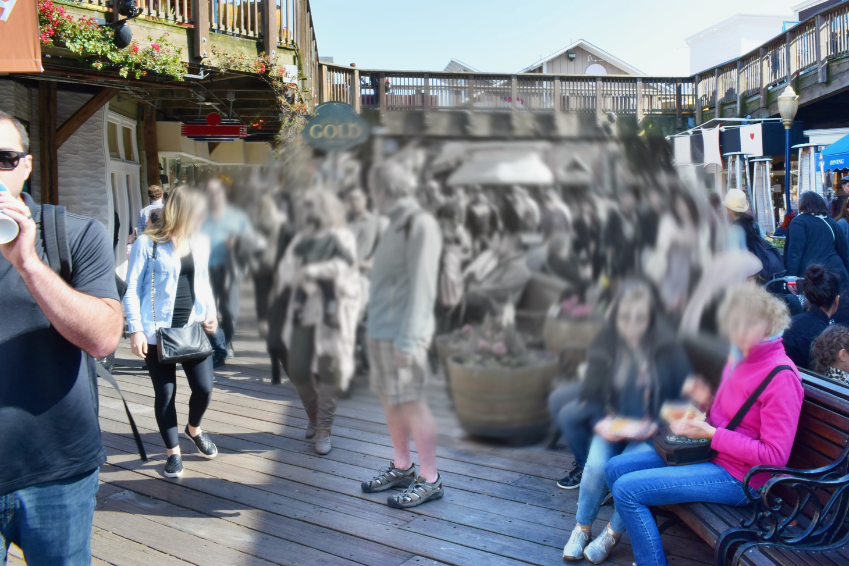
Visual field loss and scotoma in Retinal Degeneration: the variety of manifestations

Visual field loss and scotoma: Four manifestations of this Retinal Degeneration symptom can be distinguished. Tunnel vision is one of them.
Within the spectrum of visual changes in retinal degeneration, “visual field loss” proves to be a multifaceted challenge.
This Retinal Degeneration symptom represents a restriction in peripheral vision, which can manifest itself in various ways. A total of four main manifestations can be distinguished.
Scotomas: point-like or regional defects in the visual field
Scotomas are localised areas in the field of vision in which perception is impaired or completely interrupted.
These losses can appear as punctiform gaps or as larger areas in which visual information is not recognised.
Scotomas can occur in the course of retinal degeneration and impair peripheral vision.
Hemianopsia: Half-sided visual field loss
Hemianopsia describes the loss of visual perception on one half of the field of vision, be it right or left.
In right hemianopsia, the peripheral areas of the right field of vision are affected, while in left hemianopsia the corresponding areas on the left side are lost.
This form of visual field loss can significantly impair orientation and navigation in space.
Tunnel vision: Concentrated visual area with peripheral visual field loss
Tunnel vision is an impressive visual phenomenon. This is a prominent manifestation of visual field loss in which the central visual area is relatively well preserved, while the peripheral areas of the visual field are restricted or even completely lost.
This visual change can mean that those affected can literally only see through a narrow “tunnel” of visual information.
Objects and events outside this limited area can only be perceived to a limited extent or not at all.
This reduced perception of the environment can significantly impair orientation and spatial understanding, which challenges independence in everyday life.
Tunnel vision illustrates the complex nature of visual field changes in retinal degeneration.
Homonymous hemianopsia: Mirror-image visual field loss on both sides
Homonymous hemianopsia involves symmetrical losses on both sides of the visual field.
This means that a loss occurs on the right side of the field of vision in both the right and left eye, and vice versa on the left side. This form can further complicate the perception and recognition of objects.
Here are some aids that can be used for homonymous hemianopsia:
- Prism glasses: Special glasses with prism lenses can widen the field of vision by redirecting light and directing it into the impaired area.
- Mirrors: So-called “hemianopsia mirrors” can be positioned at the edge of the field of vision to reflect visual information from the affected side into the field of vision.
- Magnifying visual aids: These can facilitate the perception of objects and text by bringing them closer, thus intensifying the visual information.
- Computer software: Specialised software can expand the field of vision on the screen by transferring visual information from the affected side to the intact side.
- Eye training: Targeted eye training, exercises and therapies aimed at training the brain to process and compensate for visual information from the affected side.
- Mobility training: Training to navigate safely in the environment to recognise and avoid obstacles and dangers on the affected side.
- Advice from specialists: Specialists such as ophthalmologists, neurologists and occupational therapists can recommend individual strategies and aids to make it easier to deal with homonymous hemianopsia.
These aids can be customised to meet the needs and requirements of those affected and help them to cope with their visual impairments.
Diverse manifestations of visual field loss in Retinal Degeneration
Visual field loss in retinal degeneration takes a wide range of forms, including scotomas, hemianopsia, tunnel vision and homonymous hemianopsia.
These diverse manifestations illustrate the complexity of peripheral visual impairment.
Scotomas manifest punctate or regional losses, while hemianopsia causes hemianopic losses. Tunnel vision concentrates the central visual area with peripheral loss, and homonymous hemianopsia leads to symmetrical deficits.
Each form presents specific challenges in everyday life. Early diagnosis, targeted adaptation strategies and visual aids offer ways of coping.

